 Movie
Movie Controller

[English] 日本語
 Yorodumi
- PDB-1myw: CRYSTAL STRUCTURE OF A YELLOW FLUORESCENT PROTEIN WITH IMPROVED M... -
+ Open data

- Basic information
| Entry | Database: PDB / ID: 1myw | ||||||
|---|---|---|---|---|---|---|---|
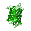
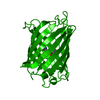
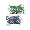
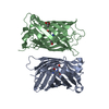
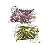
| Title | CRYSTAL STRUCTURE OF A YELLOW FLUORESCENT PROTEIN WITH IMPROVED MATURATION AND REDUCED ENVIRONMENTAL SENSITIVITY | ||||||
 Components Components | Green fluorescent protein | ||||||
 Keywords Keywords | LUMINESCENT PROTEIN / BIOLUMINESCENCE / PHOTOACTIVE PROTEIN / GREEN FLUORESCENT PROTEIN / YELLOW-EMISSION VARIANT / IMPROVED MATURATION / BETA-BARREL / CHROMOPHORE | ||||||
| Function / homology |  Function and homology information Function and homology information | ||||||
| Biological species |   Aequorea victoria (jellyfish) Aequorea victoria (jellyfish) | ||||||
| Method |  X-RAY DIFFRACTION / MOLECULAR REPLACEMENT / Resolution: 2.2 Å X-RAY DIFFRACTION / MOLECULAR REPLACEMENT / Resolution: 2.2 Å | ||||||
 Authors Authors | Rekas, A. / Alattia, J.R. / Nagai, T. / Miyawaki, A. / Ikura, M. | ||||||
 Citation Citation | Journal: J.Biol.Chem. / Year: 2002 Title: Crystal Structure of Venus, a Yellow Fluorescent Protein with Improved Maturation and Reduced Environmental Sensitivity Authors: Rekas, A. / Alattia, J.R. / Nagai, T. / Miyawaki, A. / Ikura, M. | ||||||
| History |
| ||||||
| Remark 999 | SEQUENCE RESIDUES GLY65, TYR66, AND GLY67 ARE NOT PRESENT IN THE ENTRY. INSTEAD THEY ARE REPLACED WITH CR266. |
- Structure visualization
| Structure viewer | Molecule: MolmilJmol/JSmol |
|---|
- Downloads & links
-Download
| PDBx/mmCIF format | 1myw.cif.gz | 56.2 KB | Display | PDBx/mmCIF format |
|---|---|---|---|---|
| PDB format | pdb1myw.ent.gz | 43.7 KB | Display | PDB format |
| PDBx/mmJSON format | 1myw.json.gz | Tree view | PDBx/mmJSON format | |
| Others |  Other downloads Other downloads |
-Validation report
| Summary document | 1myw_validation.pdf.gz | 374.2 KB | Display | wwPDB validaton report |
|---|---|---|---|---|
| Full document | 1myw_full_validation.pdf.gz | 377.8 KB | Display | |
| Data in XML | 1myw_validation.xml.gz | 6.8 KB | Display | |
| Data in CIF | 1myw_validation.cif.gz | 9.7 KB | Display | |
| Arichive directory | https://data.pdbj.org/pub/pdb/validation_reports/my/1mywftp://data.pdbj.org/pub/pdb/validation_reports/my/1myw | HTTPS FTP |
-Related structure data
| Related structure data |  1yfpS S: Starting model for refinement |
|---|---|
| Similar structure data |


-Links
 PDBj
PDBj

- Assembly
| Deposited unit | 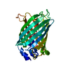
| ||||||||
|---|---|---|---|---|---|---|---|---|---|
| 1 | 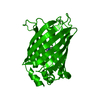
| ||||||||
| Unit cell |
|
-Components
| #1: Protein | Mass: 26875.287 Da / Num. of mol.: 1 / Fragment: residues 2-230 Mutation: F46L, F64L, S65G, V68L, S72A, M153T, V163A, S175G, T203Y Source method: isolated from a genetically manipulated source Details: SEYFP-F46L variant / Source: (gene. exp.) Aequorea victoria (jellyfish) / Plasmid: PRSET_B / Species (production host): Escherichia coli / Production host:  |
|---|---|
| #2: Water | ChemComp-HOH /  Mass: 18.015 Da / Num. of mol.: 72 / Source method: isolated from a natural source / Formula: H2O Mass: 18.015 Da / Num. of mol.: 72 / Source method: isolated from a natural source / Formula: H2O |
-Experimental details
-Experiment
| Experiment | Method: X-RAY DIFFRACTION / Number of used crystals: 1 |
|---|
- Sample preparation
| Crystal | Density Matthews: 2.65 Å3/Da / Density % sol: 53.67 % | ||||||||||||||||||||||||||||||||||||||||||||||||||||||||
|---|---|---|---|---|---|---|---|---|---|---|---|---|---|---|---|---|---|---|---|---|---|---|---|---|---|---|---|---|---|---|---|---|---|---|---|---|---|---|---|---|---|---|---|---|---|---|---|---|---|---|---|---|---|---|---|---|---|
| Crystal grow | Temperature: 298 K / Method: vapor diffusion, hanging drop / pH: 8.4 Details: Tris, ammonium sulfate, PEG400, pH 8.4, VAPOR DIFFUSION, HANGING DROP, temperature 298K | ||||||||||||||||||||||||||||||||||||||||||||||||||||||||
| Crystal grow | *PLUS pH: 7.9 | ||||||||||||||||||||||||||||||||||||||||||||||||||||||||
| Components of the solutions | *PLUS
|
-Data collection
| Diffraction | Mean temperature: 100 K |
|---|---|
| Diffraction source | Source: ROTATING ANODE / Type: RIGAKU RU200 / Wavelength: 1.5418 |
| Detector | Type: MARRESEARCH / Detector: IMAGE PLATE / Date: Nov 28, 2001 |
| Radiation | Protocol: SINGLE WAVELENGTH / Monochromatic (M) / Laue (L): M / Scattering type: x-ray |
| Radiation wavelength | Wavelength: 1.5418 Å / Relative weight: 1 |
| Reflection | Resolution: 2.2→23.88 Å / Num. all: 14560 / Num. obs: 14560 / % possible obs: 97.7 % / Redundancy: 8.34 % / Biso Wilson estimate: 21 Å2 / Rmerge(I) obs: 0.076 / Net I/σ(I): 9.2 |
| Reflection shell | Resolution: 2.2→2.34 Å / Rmerge(I) obs: 0.317 / % possible all: 94.5 |
| Reflection | *PLUS Num. obs: 121451 |
| Reflection shell | *PLUS Highest resolution: 2.28 Å / Lowest resolution: 2.37 Å |
- Processing
| Software |
| ||||||||||||||||||||||||||||||||||||
|---|---|---|---|---|---|---|---|---|---|---|---|---|---|---|---|---|---|---|---|---|---|---|---|---|---|---|---|---|---|---|---|---|---|---|---|---|---|
| Refinement | Method to determine structure: MOLECULAR REPLACEMENT Starting model: 1YFP Resolution: 2.2→23.88 Å / Rfactor Rfree error: 0.009 / Isotropic thermal model: RESTRAINED / Cross valid method: THROUGHOUT / σ(F): 0
| ||||||||||||||||||||||||||||||||||||
| Solvent computation | Solvent model: FLAT MODEL / Bsol: 52.5482 Å2 / ksol: 0.412254 e/Å3 | ||||||||||||||||||||||||||||||||||||
| Displacement parameters | Biso mean: 29.4 Å2
| ||||||||||||||||||||||||||||||||||||
| Refine analyze | Luzzati coordinate error free: 0.31 Å / Luzzati sigma a free: 0.13 Å | ||||||||||||||||||||||||||||||||||||
| Refinement step | Cycle: LAST / Resolution: 2.2→23.88 Å
| ||||||||||||||||||||||||||||||||||||
| Refine LS restraints |
| ||||||||||||||||||||||||||||||||||||
| LS refinement shell | Resolution: 2.2→2.28 Å / Rfactor Rfree error: 0.024 / Total num. of bins used: 6
| ||||||||||||||||||||||||||||||||||||
| Xplor file |
| ||||||||||||||||||||||||||||||||||||
| Refinement | *PLUS Highest resolution: 2.2 Å | ||||||||||||||||||||||||||||||||||||
| Solvent computation | *PLUS | ||||||||||||||||||||||||||||||||||||
| Displacement parameters | *PLUS | ||||||||||||||||||||||||||||||||||||
| Refine LS restraints | *PLUS
|
