 Movie
Movie Controller

[English] 日本語
 Yorodumi
- PDB-1lk7: Structure of D-Ribose-5-Phosphate Isomerase from in complex with ... -
+ Open data

- Basic information
| Entry | Database: PDB / ID: 1lk7 | ||||||
|---|---|---|---|---|---|---|---|
| Title | Structure of D-Ribose-5-Phosphate Isomerase from in complex with phospho-erythronic acid | ||||||
 Components Components | D-Ribose-5-Phosphate Isomerase | ||||||
 Keywords Keywords | ISOMERASE / alpha/beta structure | ||||||
| Function / homology |  Function and homology information Function and homology informationD-ribose metabolic process / ribose-5-phosphate isomerase / ribose-5-phosphate isomerase activity / pentose-phosphate shunt, non-oxidative branch / cytosol Similarity search - Function | ||||||
| Biological species |   Pyrococcus horikoshii (archaea) Pyrococcus horikoshii (archaea) | ||||||
| Method |  X-RAY DIFFRACTION / SYNCHROTRON / MOLECULAR REPLACEMENT / Resolution: 2 Å X-RAY DIFFRACTION / SYNCHROTRON / MOLECULAR REPLACEMENT / Resolution: 2 Å | ||||||
 Authors Authors | Ishikawa, K. / Matsui, I. / Payan, F. / Cambillau, C. / Ishida, H. / Kawarabayasi, Y. / Kikuchi, H. / Roussel, A. | ||||||
 Citation Citation | Journal: STRUCTURE / Year: 2002 Title: A Hyperthermostable D-Ribose-5-Phosphate Isomerase from Pyrococcus horikoshii Characterization and Three-Dimensional Structure Authors: Ishikawa, K. / Matsui, I. / Payan, F. / Cambillau, C. / Ishida, H. / Kawarabayasi, Y. / Kikuchi, H. / Roussel, A. | ||||||
| History |
|
- Structure visualization
| Structure viewer | Molecule: MolmilJmol/JSmol |
|---|
- Downloads & links
-Download
| PDBx/mmCIF format | 1lk7.cif.gz | 195.8 KB | Display | PDBx/mmCIF format |
|---|---|---|---|---|
| PDB format | pdb1lk7.ent.gz | 155.3 KB | Display | PDB format |
| PDBx/mmJSON format | 1lk7.json.gz | Tree view | PDBx/mmJSON format | |
| Others |  Other downloads Other downloads |
-Validation report
| Arichive directory | https://data.pdbj.org/pub/pdb/validation_reports/lk/1lk7ftp://data.pdbj.org/pub/pdb/validation_reports/lk/1lk7 | HTTPS FTP |
|---|
-Related structure data
| Related structure data |  1lk5SC S: Starting model for refinement C: citing same article ( |
|---|---|
| Similar structure data |


-Links
 PDBj
PDBj- Assembly
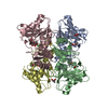
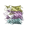
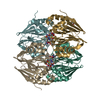
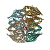
| Deposited unit | 
| ||||||||
|---|---|---|---|---|---|---|---|---|---|
| 1 |
| ||||||||
| Unit cell |
| ||||||||
| Details | The biological assembly is the tetramer in the asymetric unit |
-Components
| #1: Protein | Mass: 25189.211 Da / Num. of mol.: 4 Source method: isolated from a genetically manipulated source Source: (gene. exp.) Pyrococcus horikoshii (archaea) / Plasmid: PET11a / Species (production host): Escherichia coli / Production host:  #2: Chemical | ChemComp-CL /   Mass: 35.453 Da / Num. of mol.: 10 / Source method: obtained synthetically / Formula: Cl Mass: 35.453 Da / Num. of mol.: 10 / Source method: obtained synthetically / Formula: Cl#3: Chemical |   Mass: 22.990 Da / Num. of mol.: 2 / Source method: obtained synthetically / Formula: Na Mass: 22.990 Da / Num. of mol.: 2 / Source method: obtained synthetically / Formula: Na#4: Chemical | ChemComp-DER / 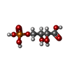  Mass: 216.083 Da / Num. of mol.: 4 / Source method: obtained synthetically / Formula: C4H9O8P Mass: 216.083 Da / Num. of mol.: 4 / Source method: obtained synthetically / Formula: C4H9O8P#5: Water | ChemComp-HOH / |  Mass: 18.015 Da / Num. of mol.: 405 / Source method: isolated from a natural source / Formula: H2O Mass: 18.015 Da / Num. of mol.: 405 / Source method: isolated from a natural source / Formula: H2O |
|---|
-Experimental details
-Experiment
| Experiment | Method: X-RAY DIFFRACTION / Number of used crystals: 1 |
|---|
- Sample preparation
| Crystal | Density Matthews: 2.51 Å3/Da / Density % sol: 51.03 % | ||||||||||||||||||||||||||||||||||||||||||
|---|---|---|---|---|---|---|---|---|---|---|---|---|---|---|---|---|---|---|---|---|---|---|---|---|---|---|---|---|---|---|---|---|---|---|---|---|---|---|---|---|---|---|---|
| Crystal grow | Temperature: 294 K / Method: vapor diffusion, hanging drop / pH: 4.6 Details: 0.1M acetate buffer, 2.0M NaCl, pH 4.6, VAPOR DIFFUSION, HANGING DROP, temperature 294K | ||||||||||||||||||||||||||||||||||||||||||
| Crystal grow | *PLUS Temperature: 20 ℃ / pH: 8 | ||||||||||||||||||||||||||||||||||||||||||
| Components of the solutions | *PLUS
|
-Data collection
| Diffraction | Mean temperature: 100 K |
|---|---|
| Diffraction source | Source: SYNCHROTRON / Site: ESRF  / Beamline: ID14-2 / Wavelength: 0.9326 Å / Beamline: ID14-2 / Wavelength: 0.9326 Å |
| Detector | Type: ADSC QUANTUM 4 / Detector: CCD |
| Radiation | Monochromator: MIRRORS / Protocol: SINGLE WAVELENGTH / Monochromatic (M) / Laue (L): M / Scattering type: x-ray |
| Radiation wavelength | Wavelength: 0.9326 Å / Relative weight: 1 |
| Reflection | Resolution: 2→15 Å / Num. all: 70244 / Num. obs: 67418 / % possible obs: 97.7 % / Observed criterion σ(F): 0 / Observed criterion σ(I): 0 |
| Reflection shell | Resolution: 2→2.03 Å / % possible all: 99.2 |
| Reflection | *PLUS Lowest resolution: 20 Å / % possible obs: 96.3 % / Redundancy: 4 % / Rmerge(I) obs: 0.05 |
| Reflection shell | *PLUS % possible obs: 96.2 % / Redundancy: 3.9 % / Rmerge(I) obs: 0.387 / Mean I/σ(I) obs: 1.9 |
- Processing
| Software |
| ||||||||||||||||||||||||
|---|---|---|---|---|---|---|---|---|---|---|---|---|---|---|---|---|---|---|---|---|---|---|---|---|---|
| Refinement | Method to determine structure: MOLECULAR REPLACEMENT Starting model: 1LK5 Resolution: 2→15 Å / Cross valid method: THROUGHOUT / σ(F): 0 / Stereochemistry target values: Engh & Huber / Details: The structure was refined also with REFMAC
| ||||||||||||||||||||||||
| Refinement step | Cycle: LAST / Resolution: 2→15 Å
| ||||||||||||||||||||||||
| Refine LS restraints |
| ||||||||||||||||||||||||
| LS refinement shell | Resolution: 2→2.051 Å
| ||||||||||||||||||||||||
| Refinement | *PLUS Num. reflection obs: 65509 / Num. reflection Rfree: 3322 / % reflection Rfree: 5 % / Rfactor Rfree: 0.27 / Rfactor Rwork: 0.239 | ||||||||||||||||||||||||
| Solvent computation | *PLUS | ||||||||||||||||||||||||
| Displacement parameters | *PLUS | ||||||||||||||||||||||||
| Refine LS restraints | *PLUS
| ||||||||||||||||||||||||
| LS refinement shell | *PLUS Rfactor Rwork: 0.25 |
