 Movie
Movie Controller

[English] 日本語
 Yorodumi
- PDB-1jpz: Crystal structure of a complex of the heme domain of P450BM-3 wit... -
+ Open data

- Basic information
| Entry | Database: PDB / ID: 1jpz | ||||||
|---|---|---|---|---|---|---|---|
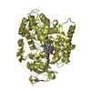
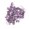
| Title | Crystal structure of a complex of the heme domain of P450BM-3 with N-Palmitoylglycine | ||||||
 Components Components | BIFUNCTIONAL P-450:NADPH-P450 REDUCTASE | ||||||
 Keywords Keywords | OXIDOREDUCTASE / Protein-substrate complex / hemeprotein | ||||||
| Function / homology |  Function and homology information Function and homology informationNADPH-hemoprotein reductase / NADPH-hemoprotein reductase activity / oxidoreductase activity, acting on paired donors, with incorporation or reduction of molecular oxygen, reduced flavin or flavoprotein as one donor, and incorporation of one atom of oxygen / unspecific monooxygenase / aromatase activity / metabolic process / response to hormone / FMN binding / flavin adenine dinucleotide binding / iron ion binding ...NADPH-hemoprotein reductase / NADPH-hemoprotein reductase activity / oxidoreductase activity, acting on paired donors, with incorporation or reduction of molecular oxygen, reduced flavin or flavoprotein as one donor, and incorporation of one atom of oxygen / unspecific monooxygenase / aromatase activity / metabolic process / response to hormone / FMN binding / flavin adenine dinucleotide binding / iron ion binding / heme binding / identical protein binding / cytosol Similarity search - Function | ||||||
| Biological species |  Bacillus megaterium (bacteria) Bacillus megaterium (bacteria) | ||||||
| Method |  X-RAY DIFFRACTION / FOURIER SYNTHESIS / Resolution: 1.65 Å X-RAY DIFFRACTION / FOURIER SYNTHESIS / Resolution: 1.65 Å | ||||||
 Authors Authors | Haines, D.C. / Tomchick, D.R. / Machius, M. / Peterson, J.A. | ||||||
 Citation Citation | Journal: Biochemistry / Year: 2001 Title: Pivotal role of water in the mechanism of P450BM-3. Authors: Haines, D.C. / Tomchick, D.R. / Machius, M. / Peterson, J.A. #1: Journal: Nat.Struct.Biol. / Year: 1997Title: The structure of the cytochrome p450BM-3 haem domain complexed with the fatty acid substrate, palmitoleic acid Authors: Li, H. / Poulos, T.L. #2: Journal: Science / Year: 2000Title: The catalytic pathway of cytochrome p450cam at atomic resolution Authors: Schlichting, I. / Berendzen, J. / Chu, K. / Stock, A.M. / Maves, S.A. / Benson, D.E. / Sweet, R.M. / Ringe, D. / Petsko, G.A. / Sligar, S.G. #3: Journal: Proc.Natl.Acad.Sci.USA / Year: 1999Title: Structure of a cytochrome P450-redox partner electron-transfer complex Authors: Sevrioukova, I.F. / Li, H. / Zang, H. / Peterson, J.A. / Poulos, T.L. | ||||||
| History |
|
- Structure visualization
| Structure viewer | Molecule: MolmilJmol/JSmol |
|---|
- Downloads & links
-Download
| PDBx/mmCIF format | 1jpz.cif.gz | 222 KB | Display | PDBx/mmCIF format |
|---|---|---|---|---|
| PDB format | pdb1jpz.ent.gz | 174 KB | Display | PDB format |
| PDBx/mmJSON format | 1jpz.json.gz | Tree view | PDBx/mmJSON format | |
| Others |  Other downloads Other downloads |
-Validation report
| Summary document | 1jpz_validation.pdf.gz | 1.4 MB | Display | wwPDB validaton report |
|---|---|---|---|---|
| Full document | 1jpz_full_validation.pdf.gz | 1.4 MB | Display | |
| Data in XML | 1jpz_validation.xml.gz | 44.6 KB | Display | |
| Data in CIF | 1jpz_validation.cif.gz | 67.1 KB | Display | |
| Arichive directory | https://data.pdbj.org/pub/pdb/validation_reports/jp/1jpzftp://data.pdbj.org/pub/pdb/validation_reports/jp/1jpz | HTTPS FTP |
-Related structure data
| Related structure data |  1fagS S: Starting model for refinement |
|---|---|
| Similar structure data |


-Links
 PDBj
PDBj


- Assembly
| Deposited unit | 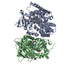
| ||||||||||
|---|---|---|---|---|---|---|---|---|---|---|---|
| 1 | 
| ||||||||||
| 2 | 
| ||||||||||
| Unit cell |
|
-Components
| #1: Protein | Mass: 53898.422 Da / Num. of mol.: 2 / Fragment: CYTOCHROME P450 102 DOMAIN Source method: isolated from a genetically manipulated source Source: (gene. exp.) Bacillus megaterium (bacteria) / Plasmid: pProEx-1 / Production host: #2: Chemical |   Mass: 616.487 Da / Num. of mol.: 2 / Source method: obtained synthetically / Formula: C34H32FeN4O4 Mass: 616.487 Da / Num. of mol.: 2 / Source method: obtained synthetically / Formula: C34H32FeN4O4#3: Chemical |   Mass: 313.475 Da / Num. of mol.: 2 / Source method: obtained synthetically / Formula: C18H35NO3 Mass: 313.475 Da / Num. of mol.: 2 / Source method: obtained synthetically / Formula: C18H35NO3#4: Water | ChemComp-HOH / |  Mass: 18.015 Da / Num. of mol.: 955 / Source method: isolated from a natural source / Formula: H2O Mass: 18.015 Da / Num. of mol.: 955 / Source method: isolated from a natural source / Formula: H2O |
|---|
-Experimental details
-Experiment
| Experiment | Method: X-RAY DIFFRACTION / Number of used crystals: 1 |
|---|
- Sample preparation
| Crystal | Density Matthews: 2.8 Å3/Da / Density % sol: 55 % | ||||||||||||||||||||||||||||||
|---|---|---|---|---|---|---|---|---|---|---|---|---|---|---|---|---|---|---|---|---|---|---|---|---|---|---|---|---|---|---|---|
| Crystal grow | Temperature: 277 K / Method: vapor diffusion, hanging drop / pH: 6 Details: 6% PEG 3350, 25 mM potassium phosphate, 50 mM magnesium chloride, 50 mM Morpholinoethanesulfonic acid pH 6.0, VAPOR DIFFUSION, HANGING DROP at 277K | ||||||||||||||||||||||||||||||
| Crystal grow | *PLUS Temperature: 4 ℃ / pH: 6 | ||||||||||||||||||||||||||||||
| Components of the solutions | *PLUS
|
-Data collection
| Diffraction | Mean temperature: 100 K |
|---|---|
| Diffraction source | Source: ROTATING ANODE / Type: RIGAKU RU200 / Wavelength: 1.5418 Å |
| Detector | Type: RIGAKU RAXIS IV / Detector: IMAGE PLATE / Date: Aug 31, 2000 |
| Radiation | Monochromator: OSMIC MIRRORS / Protocol: SINGLE WAVELENGTH / Monochromatic (M) / Laue (L): M / Scattering type: x-ray |
| Radiation wavelength | Wavelength: 1.5418 Å / Relative weight: 1 |
| Reflection | Resolution: 1.65→30 Å / Num. all: 259395 / Num. obs: 218743 / % possible obs: 84.3 % / Observed criterion σ(F): 0 / Observed criterion σ(I): 0 / Redundancy: 2.2 % / Biso Wilson estimate: 26.6 Å2 / Rmerge(I) obs: 0.031 / Net I/σ(I): 23.1 |
| Reflection shell | Resolution: 1.65→1.68 Å / Redundancy: 1.5 % / Rmerge(I) obs: 0.284 / Mean I/σ(I) obs: 1.9 / Num. unique all: 4962 / % possible all: 38.3 |
| Reflection | *PLUS Lowest resolution: 30 Å |
| Reflection shell | *PLUS % possible obs: 38.3 % |
- Processing
| Software |
| |||||||||||||||||||||||||||
|---|---|---|---|---|---|---|---|---|---|---|---|---|---|---|---|---|---|---|---|---|---|---|---|---|---|---|---|---|
| Refinement | Method to determine structure: FOURIER SYNTHESIS Starting model: PDB ENTRY 1FAG Resolution: 1.65→30 Å / Isotropic thermal model: ANISOTROPIC / Cross valid method: THROUGHOUT / σ(F): 0 / σ(I): 0 / Stereochemistry target values: ENGH & HUBER Details: THE PHI, PSI TORSION ANGLES OF LEUCINE 437 IN BOTH MONOMERS FALL IN THE DISALLOWED REGIONS, BUT THE DENSITY FOR THESE RESIDUES IS QUITE CLEAR. THEY OCCUR IN A TIGHT TURN NEAR THE SUBSTRATE BINDING CAVITY.
| |||||||||||||||||||||||||||
| Solvent computation | Bsol: 49.7 Å2 / ksol: 0.369 e/Å3 | |||||||||||||||||||||||||||
| Displacement parameters | Biso mean: 25.4 Å2
| |||||||||||||||||||||||||||
| Refine analyze | Luzzati coordinate error obs: 0.23 Å / Luzzati d res low obs: 5 Å / Luzzati sigma a obs: 0.15 Å | |||||||||||||||||||||||||||
| Refinement step | Cycle: LAST / Resolution: 1.65→30 Å
| |||||||||||||||||||||||||||
| Refine LS restraints |
| |||||||||||||||||||||||||||
| LS refinement shell | Resolution: 1.65→1.71 Å / Total num. of bins used: 10
| |||||||||||||||||||||||||||
| Xplor file |
| |||||||||||||||||||||||||||
| Software | *PLUS Name: CNS / Version: 1 / Classification: refinement | |||||||||||||||||||||||||||
| Refinement | *PLUS Lowest resolution: 30 Å / σ(F): 0 / % reflection Rfree: 4.7 % / Rfactor obs: 0.177 | |||||||||||||||||||||||||||
| Solvent computation | *PLUS | |||||||||||||||||||||||||||
| Displacement parameters | *PLUS Biso mean: 25.4 Å2 | |||||||||||||||||||||||||||
| Refine LS restraints | *PLUS
| |||||||||||||||||||||||||||
| LS refinement shell | *PLUS Rfactor Rfree: 0.474 / Rfactor Rwork: 0.439 |
