 Movie
Movie Controller

[English] 日本語
 Yorodumi
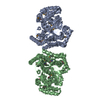
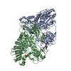
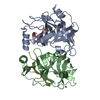
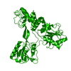
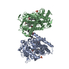
- PDB-1gx6: Hepatitis C Virus RNA polymerase in complex with UTP and manganese -
+ Open data

- Basic information
| Entry | Database: PDB / ID: 1gx6 | ||||||
|---|---|---|---|---|---|---|---|
| Title | Hepatitis C Virus RNA polymerase in complex with UTP and manganese | ||||||
 Components Components | RNA-DIRECTED RNA POLYMERASE | ||||||
 Keywords Keywords | TRANSFERASE / POLYMERASE / RNA-DEPENDENT RNA POLYMERASE / VIRUS REPLICATION / INITIATION / POLYPROTEIN / GLYCOPROTEIN / RNA-DIRECTED RNA POLYMERASE | ||||||
| Function / homology |  Function and homology information Function and homology informationhepacivirin / host cell mitochondrial membrane / host cell lipid droplet / symbiont-mediated transformation of host cell / symbiont-mediated suppression of host TRAF-mediated signal transduction / symbiont-mediated perturbation of host cell cycle G1/S transition checkpoint / symbiont-mediated suppression of host JAK-STAT cascade via inhibition of STAT1 activity / symbiont-mediated suppression of host cytoplasmic pattern recognition receptor signaling pathway via inhibition of MAVS activity / SH3 domain binding / nucleoside-triphosphate phosphatase ...hepacivirin / host cell mitochondrial membrane / host cell lipid droplet / symbiont-mediated transformation of host cell / symbiont-mediated suppression of host TRAF-mediated signal transduction / symbiont-mediated perturbation of host cell cycle G1/S transition checkpoint / symbiont-mediated suppression of host JAK-STAT cascade via inhibition of STAT1 activity / symbiont-mediated suppression of host cytoplasmic pattern recognition receptor signaling pathway via inhibition of MAVS activity / SH3 domain binding / nucleoside-triphosphate phosphatase / viral nucleocapsid / channel activity / monoatomic ion transmembrane transport / clathrin-dependent endocytosis of virus by host cell / Hydrolases; Acting on peptide bonds (peptidases); Cysteine endopeptidases / RNA helicase activity / host cell perinuclear region of cytoplasm / host cell endoplasmic reticulum membrane / RNA helicase / symbiont-mediated suppression of host type I interferon-mediated signaling pathway / ribonucleoprotein complex / serine-type endopeptidase activity / symbiont-mediated activation of host autophagy / RNA-directed RNA polymerase / cysteine-type endopeptidase activity / viral RNA genome replication / RNA-directed RNA polymerase activity / fusion of virus membrane with host endosome membrane / viral envelope / virion attachment to host cell / host cell nucleus / host cell plasma membrane / virion membrane / structural molecule activity / ATP hydrolysis activity / proteolysis / RNA binding / zinc ion binding / ATP binding Similarity search - Function | ||||||
| Biological species |  HEPATITIS C VIRUS HEPATITIS C VIRUS | ||||||
| Method |  X-RAY DIFFRACTION / SYNCHROTRON / MOLECULAR REPLACEMENT / Resolution: 1.85 Å X-RAY DIFFRACTION / SYNCHROTRON / MOLECULAR REPLACEMENT / Resolution: 1.85 Å | ||||||
 Authors Authors | Bressanelli, S. / Rey, F.A. | ||||||
 Citation Citation | Journal: J.Virol. / Year: 2002 Title: A Structural Analysis of the Hepatitis C Virus RNA Polymerase in Complex with Ribonucleotides Authors: Bressanelli, S. / Tomei, L. / Rey, F.A. / De Francesco, R. | ||||||
| History |
|
- Structure visualization
| Structure viewer | Molecule: MolmilJmol/JSmol |
|---|
- Downloads & links
-Download
| PDBx/mmCIF format | 1gx6.cif.gz | 129.7 KB | Display | PDBx/mmCIF format |
|---|---|---|---|---|
| PDB format | pdb1gx6.ent.gz | 100.5 KB | Display | PDB format |
| PDBx/mmJSON format | 1gx6.json.gz | Tree view | PDBx/mmJSON format | |
| Others |  Other downloads Other downloads |
-Validation report
| Arichive directory | https://data.pdbj.org/pub/pdb/validation_reports/gx/1gx6ftp://data.pdbj.org/pub/pdb/validation_reports/gx/1gx6 | HTTPS FTP |
|---|
-Related structure data
| Related structure data | 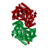 1gx5C  1csjS S: Starting model for refinement C: citing same article ( |
|---|---|
| Similar structure data |


-Links
 PDBj
PDBj
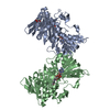
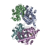
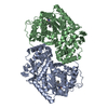
- Assembly
| Deposited unit | 
| ||||||||
|---|---|---|---|---|---|---|---|---|---|
| 1 | 
| ||||||||
| Unit cell |
| ||||||||
| Details | THE BIOLOGICAL OLIGOMERIZATION STATE OF THE PROTEIN IS NOT KNOWN.THE PARTICULAR CRYSTAL CONTACT OBSERVED IN THIS CRYSTAL IS NOTOBSERVED IN OTHER CRYSTAL FORMS OF THE PROTEIN |
-Components
| #1: Protein | Mass: 59618.484 Da / Num. of mol.: 1 / Fragment: CATALYTIC DOMAIN, RESIDUES 2420-2950 Source method: isolated from a genetically manipulated source Source: (gene. exp.) HEPATITIS C VIRUS (ISOLATE BK) / Plasmid: PT7.7 / Production host:  | ||||||
|---|---|---|---|---|---|---|---|
| #2: Chemical |   Mass: 484.141 Da / Num. of mol.: 3 / Source method: obtained synthetically / Formula: C9H15N2O15P3 / Comment: UTP*YM Mass: 484.141 Da / Num. of mol.: 3 / Source method: obtained synthetically / Formula: C9H15N2O15P3 / Comment: UTP*YM#3: Chemical | ChemComp-MN /   Mass: 54.938 Da / Num. of mol.: 5 / Source method: obtained synthetically / Formula: Mn Mass: 54.938 Da / Num. of mol.: 5 / Source method: obtained synthetically / Formula: Mn#4: Water | ChemComp-HOH / |  Mass: 18.015 Da / Num. of mol.: 485 / Source method: isolated from a natural source / Formula: H2O Mass: 18.015 Da / Num. of mol.: 485 / Source method: isolated from a natural source / Formula: H2OHas protein modification | Y | |
-Experimental details
-Experiment
| Experiment | Method: X-RAY DIFFRACTION / Number of used crystals: 1 |
|---|
- Sample preparation
| Crystal | Density Matthews: 2.6 Å3/Da / Density % sol: 51 % | ||||||||||||||||||||||||||||||||||||||||
|---|---|---|---|---|---|---|---|---|---|---|---|---|---|---|---|---|---|---|---|---|---|---|---|---|---|---|---|---|---|---|---|---|---|---|---|---|---|---|---|---|---|
| Crystal grow | pH: 6.8 Details: 4% (W/V) PEG 4000, 0.1 M SODIUM CITRATE, 7% 2-PROPANOL, pH 6.80 | ||||||||||||||||||||||||||||||||||||||||
| Crystal grow | *PLUS Temperature: 4 ℃ / pH: 7.5 / Method: vapor diffusion, hanging dropDetails: Bressanelli, S., (1999) Proc. Natl. Acad. Sci. U.S.A., 96, 13034. | ||||||||||||||||||||||||||||||||||||||||
| Components of the solutions | *PLUS
|
-Data collection
| Diffraction | Mean temperature: 100 K |
|---|---|
| Diffraction source | Source: SYNCHROTRON / Site: ESRF  / Beamline: ID14-2 / Wavelength: 0.9326 / Beamline: ID14-2 / Wavelength: 0.9326 |
| Detector | Type: ADSC CCD / Detector: CCD / Date: Sep 15, 2000 |
| Radiation | Protocol: SINGLE WAVELENGTH / Monochromatic (M) / Laue (L): M / Scattering type: x-ray |
| Radiation wavelength | Wavelength: 0.9326 Å / Relative weight: 1 |
| Reflection | Resolution: 1.85→25 Å / Num. obs: 53875 / % possible obs: 99.5 % / Observed criterion σ(I): -3 / Redundancy: 3.5 % / Biso Wilson estimate: 23.5 Å2 / Rmerge(I) obs: 0.075 / Net I/σ(I): 12.3 |
| Reflection shell | Resolution: 1.85→1.88 Å / Redundancy: 3.4 % / Rmerge(I) obs: 0.384 / Mean I/σ(I) obs: 3.3 / % possible all: 99.9 |
| Reflection | *PLUS Lowest resolution: 25 Å |
| Reflection shell | *PLUS % possible obs: 99.9 % |
- Processing
| Software |
| ||||||||||||||||||||||||||||||||||||||||||||||||||||||||||||||||||||||||||||||||
|---|---|---|---|---|---|---|---|---|---|---|---|---|---|---|---|---|---|---|---|---|---|---|---|---|---|---|---|---|---|---|---|---|---|---|---|---|---|---|---|---|---|---|---|---|---|---|---|---|---|---|---|---|---|---|---|---|---|---|---|---|---|---|---|---|---|---|---|---|---|---|---|---|---|---|---|---|---|---|---|---|---|
| Refinement | Method to determine structure: MOLECULAR REPLACEMENT Starting model: PDB ENTRY 1CSJ Resolution: 1.85→25 Å / Rfactor Rfree error: 0.004 / Data cutoff high absF: 1900318.67 / Isotropic thermal model: RESTRAINED / Cross valid method: THROUGHOUT / σ(F): 0 / Stereochemistry target values: MAXIMUM LIKELIHOOD Details: BULK SOLVENT MODEL USED 5 C-TERMINAL RESIDUES DISORDERED AND NOT INCLUDED IN MODEL
| ||||||||||||||||||||||||||||||||||||||||||||||||||||||||||||||||||||||||||||||||
| Solvent computation | Solvent model: FLAT MODEL / Bsol: 48.2094 Å2 / ksol: 0.316111 e/Å3 | ||||||||||||||||||||||||||||||||||||||||||||||||||||||||||||||||||||||||||||||||
| Displacement parameters | Biso mean: 36.5 Å2
| ||||||||||||||||||||||||||||||||||||||||||||||||||||||||||||||||||||||||||||||||
| Refine analyze |
| ||||||||||||||||||||||||||||||||||||||||||||||||||||||||||||||||||||||||||||||||
| Refinement step | Cycle: LAST / Resolution: 1.85→25 Å
| ||||||||||||||||||||||||||||||||||||||||||||||||||||||||||||||||||||||||||||||||
| Refine LS restraints |
| ||||||||||||||||||||||||||||||||||||||||||||||||||||||||||||||||||||||||||||||||
| LS refinement shell | Resolution: 1.85→1.88 Å / Rfactor Rfree error: 0.031 / Total num. of bins used: 20
| ||||||||||||||||||||||||||||||||||||||||||||||||||||||||||||||||||||||||||||||||
| Xplor file |
| ||||||||||||||||||||||||||||||||||||||||||||||||||||||||||||||||||||||||||||||||
| Refinement | *PLUS Lowest resolution: 25 Å / % reflection Rfree: 5 % | ||||||||||||||||||||||||||||||||||||||||||||||||||||||||||||||||||||||||||||||||
| Solvent computation | *PLUS | ||||||||||||||||||||||||||||||||||||||||||||||||||||||||||||||||||||||||||||||||
| Displacement parameters | *PLUS | ||||||||||||||||||||||||||||||||||||||||||||||||||||||||||||||||||||||||||||||||
| Refine LS restraints | *PLUS
|
