Group: Atomic model / Data collection ...Atomic model / Data collection / Database references / Derived calculations / Non-polymer description / Other / Refinement description / Source and taxonomy / Structure summary / Version format compliance
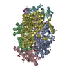
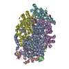
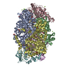
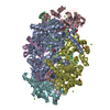
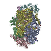
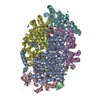
A: METHYL-COENZYME M REDUCTASE SUBUNIT ALPHA B: METHYL-COENZYME M REDUCTASE I BETA SUBUNIT C: METHYL-COENZYME M REDUCTASE SUBUNIT GAMMA D: METHYL-COENZYME M REDUCTASE SUBUNIT ALPHA E: METHYL-COENZYME M REDUCTASE I BETA SUBUNIT F: METHYL-COENZYME M REDUCTASE SUBUNIT GAMMA hetero molecules
Mass: 18.015 Da / Num. of mol.: 2195 / Source method: isolated from a natural source / Formula: H2O
-
Details
Compound details
THE HEXAMER OF TWO ALPHA, TWO BETA, AND TWO GAMMA CHAINS CATALYZES THE FINAL STEP IN ...THE HEXAMER OF TWO ALPHA, TWO BETA, AND TWO GAMMA CHAINS CATALYZES THE FINAL STEP IN METHANOGENESIS, WHICH IS THE TERMINAL STEP OF ANAEROBIC DEGRADATION OF BIOMASS
-
Experimental details
-
Experiment
Experiment
Method: X-RAY DIFFRACTION / Number of used crystals: 1
-
Sample preparation
Crystal
Density Matthews: 2.47 Å3/Da / Density % sol: 39 %
Crystal grow
pH: 7 / Details: pH 7.00
Crystal grow
*PLUS
Temperature: 14 ℃ / Method: vapor diffusion, hanging drop
In the structure databanks used in Yorodumi, some data are registered as the other names, "COVID-19 virus" and "2019-nCoV". Here are the details of the virus and the list of structure data.
Jan 31, 2019. EMDB accession codes are about to change! (news from PDBe EMDB page)
EMDB accession codes are about to change! (news from PDBe EMDB page)
The allocation of 4 digits for EMDB accession codes will soon come to an end. Whilst these codes will remain in use, new EMDB accession codes will include an additional digit and will expand incrementally as the available range of codes is exhausted. The current 4-digit format prefixed with “EMD-” (i.e. EMD-XXXX) will advance to a 5-digit format (i.e. EMD-XXXXX), and so on. It is currently estimated that the 4-digit codes will be depleted around Spring 2019, at which point the 5-digit format will come into force.
The EM Navigator/Yorodumi systems omit the EMD- prefix.
Related info.:Q: What is EMD? / ID/Accession-code notation in Yorodumi/EM Navigator
Yorodumi is a browser for structure data from EMDB, PDB, SASBDB, etc.
This page is also the successor to EM Navigator detail page, and also detail information page/front-end page for Omokage search.
The word "yorodu" (or yorozu) is an old Japanese word meaning "ten thousand". "mi" (miru) is to see.
Related info.:EMDB / PDB / SASBDB / Comparison of 3 databanks / Yorodumi Search / Aug 31, 2016. New EM Navigator & Yorodumi / Yorodumi Papers / Jmol/JSmol / Function and homology information / Changes in new EM Navigator and Yorodumi
 Movie
Movie Controller


 Open data
 Basic information
 Components
 Keywords
 Function and homology information
 METHANOSARCINA BARKERI (archaea)
 X-RAY DIFFRACTION /
  Authors
 Citation
 Structure visualization
 Downloads & links
 Other downloads
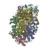


 PDBj
PDBj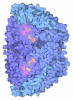
 Assembly


 Mass: 906.580 Da / Num. of mol.: 2 / Source method: obtained synthetically / Formula: C42H51N6NiO13
 Mass: 343.334 Da / Num. of mol.: 2 / Source method: obtained synthetically / Formula: C11H22NO7PS
 Mass: 142.197 Da / Num. of mol.: 2 / Source method: obtained synthetically / Formula: C2H6O3S2
 Mass: 92.094 Da / Num. of mol.: 3 / Source method: obtained synthetically / Formula: C3H8O3
 Sample preparation
 / Beamline: BW7B / Wavelength: 0.84
  Processing