[English] 日本語
 Yorodumi
- PDB-1d6y: CRYSTAL STRUCTURE OF E. COLI COPPER-CONTAINING AMINE OXIDASE ANAE... -
+ Open data
- Basic information
| Entry | Database: PDB / ID: 1d6y | ||||||
|---|---|---|---|---|---|---|---|
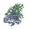
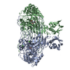
| Title | CRYSTAL STRUCTURE OF E. COLI COPPER-CONTAINING AMINE OXIDASE ANAEROBICALLY REDUCED WITH BETA-PHENYLETHYLAMINE AND COMPLEXED WITH NITRIC OXIDE. | ||||||
 Components Components | COPPER AMINE OXIDASE | ||||||
 Keywords Keywords | OXIDOREDUCTASE / REACTION INTERMEDIATE MIMIC | ||||||
| Function / homology |  Function and homology information Function and homology informationphenylethylamine catabolic process / primary-amine oxidase / primary methylamine oxidase activity / amine metabolic process / L-phenylalanine catabolic process / quinone binding / periplasmic space / copper ion binding / calcium ion binding Similarity search - Function | ||||||
| Biological species |  | ||||||
| Method |  X-RAY DIFFRACTION / X-RAY DIFFRACTION /  SYNCHROTRON / Resolution: 2.4 Å SYNCHROTRON / Resolution: 2.4 Å | ||||||
 Authors Authors | Wilmot, C.M. / Hajdu, J. / McPherson, M.J. / Knowles, P.F. / Phillips, S.E.V. | ||||||
 Citation Citation |  Journal: Science / Year: 1999 Journal: Science / Year: 1999Title: Visualization of dioxygen bound to copper during enzyme catalysis. Authors: Wilmot, C.M. / Hajdu, J. / McPherson, M.J. / Knowles, P.F. / Phillips, S.E. #1:  Journal: Biochemistry / Year: 1999 Journal: Biochemistry / Year: 1999Title: The Active Site Base Controls Cofactor Reactivity in Escherichia coli Amine Oxidase: X-ray Crystallographic Studies with Mutational Variants. Authors: Murray, J.M. / Saysell, C.G. / Wilmot, C.M. / Tambyrajah, W.S. / Jaeger, J. / Knowles, P.F. / Phillips, S.E. / McPherson, M.J. #2:  Journal: Biochemistry / Year: 1997 Journal: Biochemistry / Year: 1997Title: Catalytic Mechanism of the Quinoenzyme Amine Oxidase from Escherichia coli: Exploring the Reductive Half-Reaction. Authors: Wilmot, C.M. / Murray, J.M. / Alton, G. / Parsons, M.R. / Convery, M.A. / Blakeley, V. / Corner, A.S. / Palcic, M.M. / Knowles, P.F. / McPherson, M.J. / Knowles, P.F. #3:  Journal: Structure / Year: 1995 Journal: Structure / Year: 1995Title: Crystal Structure of a Quinoenzyme: Copper Amine Oxidase of Escherichia coli at 2 Angstroms Resolution. Authors: Parsons, M.R. / Convery, M.A. / Wilmot, C.M. / Yadav, K.D.S. / Blakeley, V. / Corner, A.S. / Phillips, S.E. / McPherson, M.J. / Knowles, P.F. | ||||||
| History |
|
- Structure visualization
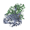
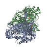
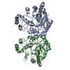
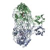
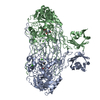
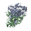
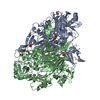
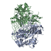
| Structure viewer | Molecule:  Molmil Molmil Jmol/JSmol Jmol/JSmol |
|---|
- Downloads & links
- Download
| PDBx/mmCIF format |  1d6y.cif.gz 1d6y.cif.gz | 327.3 KB | Display |  PDBx/mmCIF format PDBx/mmCIF format |
|---|---|---|---|---|
| PDB format |  pdb1d6y.ent.gz pdb1d6y.ent.gz | 261 KB | Display |  PDB format PDB format |
| PDBx/mmJSON format |  1d6y.json.gz 1d6y.json.gz | Tree view |  PDBx/mmJSON format PDBx/mmJSON format | |
| Others |  Other downloads Other downloads |
-Validation report
| Arichive directory |  https://data.pdbj.org/pub/pdb/validation_reports/d6/1d6y https://data.pdbj.org/pub/pdb/validation_reports/d6/1d6y ftp://data.pdbj.org/pub/pdb/validation_reports/d6/1d6y ftp://data.pdbj.org/pub/pdb/validation_reports/d6/1d6y | HTTPS FTP |
|---|
-Related structure data
- Links
Links
- Assembly
| Deposited unit | 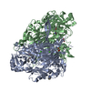
| ||||||||
|---|---|---|---|---|---|---|---|---|---|
| 1 |
| ||||||||
| Unit cell |
|
- Components
-Protein , 1 types, 2 molecules AB
| #1: Protein | Mass: 81367.758 Da / Num. of mol.: 2 Source method: isolated from a genetically manipulated source Source: (gene. exp.)   |
|---|
-Non-polymers , 7 types, 1440 molecules 



| #2: Chemical | | #3: Chemical | ChemComp-CA / #4: Chemical | #5: Chemical | #6: Chemical | ChemComp-PEA / | #7: Chemical | #8: Water | ChemComp-HOH / | |
|---|
-Details
| Has protein modification | Y |
|---|
-Experimental details
-Experiment
| Experiment | Method:  X-RAY DIFFRACTION / Number of used crystals: 1 X-RAY DIFFRACTION / Number of used crystals: 1 |
|---|
- Sample preparation
| Crystal | Density Matthews: 2.75 Å3/Da / Density % sol: 55.31 % | |||||||||||||||
|---|---|---|---|---|---|---|---|---|---|---|---|---|---|---|---|---|
| Crystal grow | Temperature: 291 K / Method: vapor diffusion, sitting drop / pH: 7.2 Details: sodium citrate, HEPES buffer, pH 7.2, VAPOR DIFFUSION, SITTING DROP, temperature 18K | |||||||||||||||
| Crystal | *PLUS Density % sol: 55 % | |||||||||||||||
| Crystal grow | *PLUS pH: 7 / Method: vapor diffusion | |||||||||||||||
| Components of the solutions | *PLUS
|
-Data collection
| Diffraction | Mean temperature: 100 K |
|---|---|
| Diffraction source | Source:  SYNCHROTRON / Site: SYNCHROTRON / Site:  ESRF ESRF  / Beamline: BM14 / Wavelength: 1.03 / Beamline: BM14 / Wavelength: 1.03 |
| Detector | Type: MARRESEARCH / Detector: IMAGE PLATE / Date: May 6, 1998 |
| Radiation | Protocol: SINGLE WAVELENGTH / Monochromatic (M) / Laue (L): M / Scattering type: x-ray |
| Radiation wavelength | Wavelength: 1.03 Å / Relative weight: 1 |
| Reflection | Resolution: 2.4→30 Å / Num. all: 65769 / Num. obs: 191118 / % possible obs: 92.5 % / Observed criterion σ(F): 0 / Observed criterion σ(I): 0 / Redundancy: 2.9 % / Biso Wilson estimate: 46.1 Å2 / Rmerge(I) obs: 0.074 / Net I/σ(I): 16 |
| Reflection shell | Resolution: 2.4→2.44 Å / Redundancy: 3.6 % / Rmerge(I) obs: 0.265 / % possible all: 92.5 |
| Reflection | *PLUS Num. obs: 65769 / Num. measured all: 191118 |
| Reflection shell | *PLUS % possible obs: 92.5 % |
- Processing
| Software |
| ||||||||||||||||||||
|---|---|---|---|---|---|---|---|---|---|---|---|---|---|---|---|---|---|---|---|---|---|
| Refinement | Resolution: 2.4→30 Å / σ(F): 0 / σ(I): 0 / Stereochemistry target values: Engh and Huber
| ||||||||||||||||||||
| Refinement step | Cycle: LAST / Resolution: 2.4→30 Å
| ||||||||||||||||||||
| Refine LS restraints |
| ||||||||||||||||||||
| Software | *PLUS Name: 'CNS' / Classification: refinement | ||||||||||||||||||||
| Refinement | *PLUS Rfactor obs: 0.181 | ||||||||||||||||||||
| Solvent computation | *PLUS | ||||||||||||||||||||
| Displacement parameters | *PLUS |
 Movie
Movie Controller








 PDBj
PDBj


