- EMDB-1285: Structure of the ribosome-bound cricket paralysis virus IRES RNA. -
+
Open data
ID or keywords:
Loading...
-
Basic information
Entry
Database: EMDB / ID: EMD-1285
Title
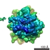
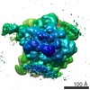
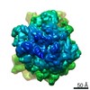
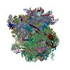
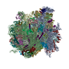
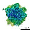
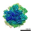
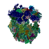
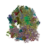
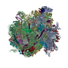
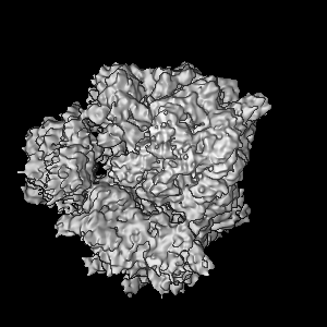
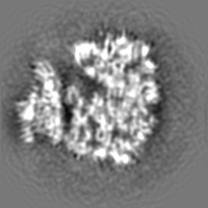
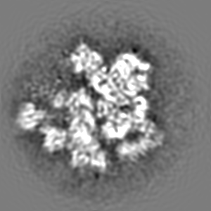
Structure of the ribosome-bound cricket paralysis virus IRES RNA.
Map data
Complex between the yeast 80S ribosome and the cricket paralysis virus IRES
Sample
Sample: yeast 80S ribosome in complex with the CrPV IRES RNA
Complex: yeast 80S ribosome
RNA: CrPV IRES
Function / homology
Function and homology information
Formation of the ternary complex, and subsequently, the 43S complex / Translation initiation complex formation / Ribosomal scanning and start codon recognition / SRP-dependent cotranslational protein targeting to membrane / GTP hydrolysis and joining of the 60S ribosomal subunit / Formation of a pool of free 40S subunits / Nonsense Mediated Decay (NMD) independent of the Exon Junction Complex (EJC) / Nonsense Mediated Decay (NMD) enhanced by the Exon Junction Complex (EJC) / L13a-mediated translational silencing of Ceruloplasmin expression / ribosomal large subunit export from nucleus ...Formation of the ternary complex, and subsequently, the 43S complex / Translation initiation complex formation / Ribosomal scanning and start codon recognition / SRP-dependent cotranslational protein targeting to membrane / GTP hydrolysis and joining of the 60S ribosomal subunit / Formation of a pool of free 40S subunits / Nonsense Mediated Decay (NMD) independent of the Exon Junction Complex (EJC) / Nonsense Mediated Decay (NMD) enhanced by the Exon Junction Complex (EJC) / L13a-mediated translational silencing of Ceruloplasmin expression / ribosomal large subunit export from nucleus / 90S preribosome / ribosomal subunit export from nucleus / regulation of translational fidelity / maturation of LSU-rRNA / maturation of SSU-rRNA / ribosomal large subunit assembly / cytosolic small ribosomal subunit / cytosolic large ribosomal subunit / cytoplasmic translation / rRNA binding / structural constituent of ribosome / ribosome / translation / mRNA binding / RNA binding / nucleus / cytoplasm / cytosol Similarity search - Function
Ribosomal protein L1, conserved site / Ribosomal protein L1 signature. / Ribosomal protein L1 / Ribosomal protein L1, 3-layer alpha/beta-sandwich / Ribosomal protein L1-like / Ribosomal protein L1/ribosomal biogenesis protein / Ribosomal protein L1p/L10e family / : / Ribosomal protein S5/S7, eukaryotic/archaeal / : ...Ribosomal protein L1, conserved site / Ribosomal protein L1 signature. / Ribosomal protein L1 / Ribosomal protein L1, 3-layer alpha/beta-sandwich / Ribosomal protein L1-like / Ribosomal protein L1/ribosomal biogenesis protein / Ribosomal protein L1p/L10e family / : / Ribosomal protein S5/S7, eukaryotic/archaeal / : / Ribosomal protein L5, conserved site / Ribosomal protein L5 signature. / Ribosomal protein L5, N-terminal / Ribosomal protein L5 / Ribosomal protein L5, C-terminal / ribosomal L5P family C-terminus / Ribosomal protein L5 / Ribosomal protein L5 domain superfamily / Ribosomal protein S7, conserved site / Ribosomal protein S7 signature. / Ribosomal protein S5/S7 / Ribosomal protein S7 domain / Ribosomal protein S7 domain superfamily / Ribosomal protein S7p/S5e Similarity search - Domain/homology
Large ribosomal subunit protein uL1A / Small ribosomal subunit protein uS7 / 60S ribosomal protein L1-B / Large ribosomal subunit protein uL5B Similarity search - Component
Biological species
Saccharomyces cerevisiae (brewer's yeast)
Method
single particle reconstruction / cryo EM / Resolution: 7.3 Å
Journal: Nat Struct Mol Biol / Year: 2006 Title: Structure of the ribosome-bound cricket paralysis virus IRES RNA. Authors: Martin Schüler / Sean R Connell / Aurelie Lescoute / Jan Giesebrecht / Marylena Dabrowski / Birgit Schroeer / Thorsten Mielke / Pawel A Penczek / Eric Westhof / Christian M T Spahn / Abstract: Internal ribosome entry sites (IRESs) facilitate an alternative, end-independent pathway of translation initiation. A particular family of dicistroviral IRESs can assemble elongation-competent 80S ...Internal ribosome entry sites (IRESs) facilitate an alternative, end-independent pathway of translation initiation. A particular family of dicistroviral IRESs can assemble elongation-competent 80S ribosomal complexes in the absence of canonical initiation factors and initiator transfer RNA. We present here a cryo-EM reconstruction of a dicistroviral IRES bound to the 80S ribosome. The resolution of the cryo-EM reconstruction, in the subnanometer range, allowed the molecular structure of the complete IRES in its active, ribosome-bound state to be solved. The structure, harboring three pseudoknot-containing domains, each with a specific functional role, shows how defined elements of the IRES emerge from a compactly folded core and interact with the key ribosomal components that form the A, P and E sites, where tRNAs normally bind. Our results exemplify the molecular strategy for recruitment of an IRES and reveal the dynamic features necessary for internal initiation.
History
Deposition
Oct 31, 2006
-
Header (metadata) release
Oct 31, 2006
-
Map release
Oct 31, 2007
-
Update
Oct 24, 2012
-
Current status
Oct 24, 2012
Processing site: PDBe / Status: Released
-
Structure visualization
Movie
Surface view with section colored by density value
Category: FILM / Film or detector model: KODAK SO-163 FILM / Digitization - Scanner: PRIMESCAN / Digitization - Sampling interval: 4.7 µm / Number real images: 341 / Average electron dose: 20 e/Å2
Electron beam
Acceleration voltage: 300 kV / Electron source: FIELD EMISSION GUN
In the structure databanks used in Yorodumi, some data are registered as the other names, "COVID-19 virus" and "2019-nCoV". Here are the details of the virus and the list of structure data.
Jan 31, 2019. EMDB accession codes are about to change! (news from PDBe EMDB page)
EMDB accession codes are about to change! (news from PDBe EMDB page)
The allocation of 4 digits for EMDB accession codes will soon come to an end. Whilst these codes will remain in use, new EMDB accession codes will include an additional digit and will expand incrementally as the available range of codes is exhausted. The current 4-digit format prefixed with “EMD-” (i.e. EMD-XXXX) will advance to a 5-digit format (i.e. EMD-XXXXX), and so on. It is currently estimated that the 4-digit codes will be depleted around Spring 2019, at which point the 5-digit format will come into force.
The EM Navigator/Yorodumi systems omit the EMD- prefix.
Related info.:Q: What is EMD? / ID/Accession-code notation in Yorodumi/EM Navigator
Yorodumi is a browser for structure data from EMDB, PDB, SASBDB, etc.
This page is also the successor to EM Navigator detail page, and also detail information page/front-end page for Omokage search.
The word "yorodu" (or yorozu) is an old Japanese word meaning "ten thousand". "mi" (miru) is to see.
Related info.:EMDB / PDB / SASBDB / Comparison of 3 databanks / Yorodumi Search / Aug 31, 2016. New EM Navigator & Yorodumi / Yorodumi Papers / Jmol/JSmol / Function and homology information / Changes in new EM Navigator and Yorodumi
 Movie
Movie Controller

 Yorodumi
 Open data
 Basic information
 Map data
 Sample
 Function and homology information
 Authors
 Citation
 Structure visualization
 Downloads & links
 1285.gif
 http://ftp.pdbj.org/pub/emdb/structures/EMD-1285
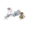


 Z (Sec.)
 Y (Row.)
 X (Col.)


 Sample components
 Processing
 Electron microscopy
 FIELD EMISSION GUN
