 Movie
Movie Controller


+ Open data

- Basic information
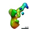
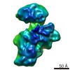
| Entry | Database: PDB / ID: 2noq | ||||||
|---|---|---|---|---|---|---|---|
| Title | Structure of ribosome-bound cricket paralysis virus IRES RNA | ||||||
 Components Components |
| ||||||
 Keywords Keywords | RIBOSOME / IRES RNA / Translation / Internal Initiation | ||||||
| Function / homology |  Function and homology information Function and homology informationRibosome Quality Control (RQC) complex extracts and degrades nascent peptide / PELO:HBS1L and ABCE1 dissociate a ribosome on a non-stop mRNA / Formation of the ternary complex, and subsequently, the 43S complex / Translation initiation complex formation / Ribosomal scanning and start codon recognition / SRP-dependent cotranslational protein targeting to membrane / GTP hydrolysis and joining of the 60S ribosomal subunit / Formation of a pool of free 40S subunits / Nonsense Mediated Decay (NMD) independent of the Exon Junction Complex (EJC) / Nonsense Mediated Decay (NMD) enhanced by the Exon Junction Complex (EJC) ...Ribosome Quality Control (RQC) complex extracts and degrades nascent peptide / PELO:HBS1L and ABCE1 dissociate a ribosome on a non-stop mRNA / Formation of the ternary complex, and subsequently, the 43S complex / Translation initiation complex formation / Ribosomal scanning and start codon recognition / SRP-dependent cotranslational protein targeting to membrane / GTP hydrolysis and joining of the 60S ribosomal subunit / Formation of a pool of free 40S subunits / Nonsense Mediated Decay (NMD) independent of the Exon Junction Complex (EJC) / Nonsense Mediated Decay (NMD) enhanced by the Exon Junction Complex (EJC) / L13a-mediated translational silencing of Ceruloplasmin expression / ribosomal large subunit export from nucleus / 90S preribosome / ribosomal subunit export from nucleus / regulation of translational fidelity / maturation of LSU-rRNA / maturation of SSU-rRNA / ribosomal large subunit assembly / cytosolic small ribosomal subunit / cytosolic large ribosomal subunit / cytoplasmic translation / rRNA binding / structural constituent of ribosome / ribosome / translation / mRNA binding / RNA binding / nucleus / cytosol / cytoplasm Similarity search - Function | ||||||
| Biological species |  | ||||||
| Method | ELECTRON MICROSCOPY / single particle reconstruction / cryo EM / Resolution: 7.3 Å | ||||||
 Authors Authors | Schuler, M. / Connell, S.R. / Lescoute, A. / Giesebrecht, J. / Dabrowski, M. / Schroeer, B. / Mielke, T. / Penczek, P.A. / Westhof, E. / Spahn, C.M.T. | ||||||
 Citation Citation | Journal: Nat Struct Mol Biol / Year: 2006 Title: Structure of the ribosome-bound cricket paralysis virus IRES RNA. Authors: Martin Schüler / Sean R Connell / Aurelie Lescoute / Jan Giesebrecht / Marylena Dabrowski / Birgit Schroeer / Thorsten Mielke / Pawel A Penczek / Eric Westhof / Christian M T Spahn /  Abstract: Internal ribosome entry sites (IRESs) facilitate an alternative, end-independent pathway of translation initiation. A particular family of dicistroviral IRESs can assemble elongation-competent 80S ...Internal ribosome entry sites (IRESs) facilitate an alternative, end-independent pathway of translation initiation. A particular family of dicistroviral IRESs can assemble elongation-competent 80S ribosomal complexes in the absence of canonical initiation factors and initiator transfer RNA. We present here a cryo-EM reconstruction of a dicistroviral IRES bound to the 80S ribosome. The resolution of the cryo-EM reconstruction, in the subnanometer range, allowed the molecular structure of the complete IRES in its active, ribosome-bound state to be solved. The structure, harboring three pseudoknot-containing domains, each with a specific functional role, shows how defined elements of the IRES emerge from a compactly folded core and interact with the key ribosomal components that form the A, P and E sites, where tRNAs normally bind. Our results exemplify the molecular strategy for recruitment of an IRES and reveal the dynamic features necessary for internal initiation. | ||||||
| History |
|
- Structure visualization
| Movie |
Movie viewer |
|---|---|
| Structure viewer | Molecule: MolmilJmol/JSmol |
- Downloads & links
-Download
| PDBx/mmCIF format | 2noq.cif.gz | 268 KB | Display | PDBx/mmCIF format |
|---|---|---|---|---|
| PDB format | pdb2noq.ent.gz | 196.7 KB | Display | PDB format |
| PDBx/mmJSON format | 2noq.json.gz | Tree view | PDBx/mmJSON format | |
| Others |  Other downloads Other downloads |
-Validation report
| Arichive directory | https://data.pdbj.org/pub/pdb/validation_reports/no/2noqftp://data.pdbj.org/pub/pdb/validation_reports/no/2noq | HTTPS FTP |
|---|
-Related structure data
| Related structure data | 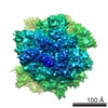 1285MC M: map data used to model this data C: citing same article ( |
|---|---|
| Similar structure data |
-Links
 PDBj
PDBj


- Assembly
| Deposited unit | 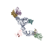
|
|---|---|
| 1 |
|
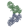
-Components
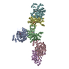
-RNA chain , 2 types, 2 molecules AE
| #1: RNA chain | Mass: 60828.805 Da / Num. of mol.: 1 / Source method: obtained synthetically Details: This chain, CrPV-IRES-RNA [NCBI accession: BD177018], was synthesized by in-vitro transcription of a plasmid template References: GenBank: 8895506 |
|---|---|
| #5: RNA chain | Mass: 17186.295 Da / Num. of mol.: 1 / Source method: isolated from a natural source / Source: (natural) |
-18S ribosomal ... , 3 types, 3 molecules BCD
| #2: RNA chain | Mass: 14869.938 Da / Num. of mol.: 1 / Fragment: residues 500-545 / Source method: isolated from a natural source / Source: (natural) |
|---|---|
| #3: RNA chain | Mass: 4149.502 Da / Num. of mol.: 1 / Fragment: residues 1050-1062 / Source method: isolated from a natural source / Source: (natural) |
| #4: RNA chain | Mass: 4784.873 Da / Num. of mol.: 1 / Fragment: residues 1194-1208 / Source method: isolated from a natural source / Source: (natural) |
-Protein , 1 types, 1 molecules F
| #6: Protein | Mass: 16577.156 Da / Num. of mol.: 1 / Source method: isolated from a natural source / Source: (natural) |
|---|
-60S ribosomal protein ... , 2 types, 2 molecules GH
| #7: Protein | Mass: 24014.168 Da / Num. of mol.: 1 / Source method: isolated from a natural source / Source: (natural) |
|---|---|
| #8: Protein | Mass: 18780.525 Da / Num. of mol.: 1 / Source method: isolated from a natural source / Source: (natural) |
-Experimental details
-Experiment
| Experiment | Method: ELECTRON MICROSCOPY |
|---|---|
| EM experiment | Aggregation state: PARTICLE / 3D reconstruction method: single particle reconstruction |
- Sample preparation
| Component | Name: Yeast 80S bound by cricket paralysis virus IRES RNA / Type: VIRUS |
|---|---|
| Buffer solution | Name: 20 mM Hepes pH 7.6, 100 mM KAc, 100 mM KCl, 5 mM MgCl2, 1 mM DTT, 1mM AEBSF, Roche Complete Protease Inhibitor pH: 7.6 Details: 20 mM Hepes pH 7.6, 100 mM KAc, 100 mM KCl, 5 mM MgCl2, 1 mM DTT, 1mM AEBSF, Roche Complete Protease Inhibitor |
| Specimen | Embedding applied: NO / Shadowing applied: NO / Staining applied: NO / Vitrification applied: YES |
| Specimen support | Details: Quantifoil |
| Vitrification | Instrument: FEI VITROBOT MARK I / Cryogen name: ETHANE |
- Electron microscopy imaging
| Experimental equipment |  Model: Tecnai Polara / Image courtesy: FEI Company |
|---|---|
| Microscopy | Model: FEI POLARA 300 |
| Electron gun | Electron source:  FIELD EMISSION GUN / Accelerating voltage: 300 kV / Illumination mode: FLOOD BEAM FIELD EMISSION GUN / Accelerating voltage: 300 kV / Illumination mode: FLOOD BEAM |
| Electron lens | Mode: BRIGHT FIELD / Nominal magnification: 39000 X / Nominal defocus max: 3900 nm / Nominal defocus min: 100 nm |
| Image recording | Electron dose: 20 e/Å2 / Film or detector model: KODAK SO-163 FILM |
| Detector | Type: KODAK SO163 FILM |
| Radiation | Protocol: SINGLE WAVELENGTH / Monochromatic (M) / Laue (L): M / Scattering type: x-ray |
| Radiation wavelength | Relative weight: 1 |
- Processing
| Symmetry | Point symmetry: C1 (asymmetric) | |||||||||||||||||||||
|---|---|---|---|---|---|---|---|---|---|---|---|---|---|---|---|---|---|---|---|---|---|---|
| 3D reconstruction | Method: Iterative 3D-Projection Matching / Resolution: 7.3 Å / Num. of particles: 73313 / Nominal pixel size: 1.22 Å / Symmetry type: POINT | |||||||||||||||||||||
| Atomic model building |
| |||||||||||||||||||||
| Refinement step | Cycle: LAST
|

