- EMDB-12237: Subtomogram average reconstruction of humanVPS34 complex II bound... -
+
Open data
ID or keywords:
Loading...
-
Basic information
Entry
Database: EMDB / ID: EMD-12237
Title
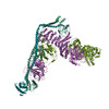
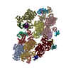
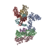
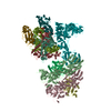
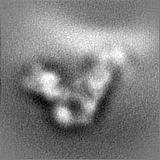
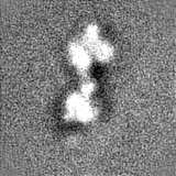
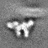
Subtomogram average reconstruction of humanVPS34 complex II bound to Rab5a on a lipid membrane
Map data
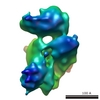
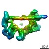
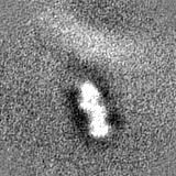
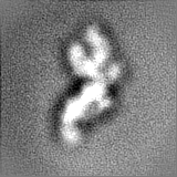
A reconstruction of the VPS34 complex II bound to Rab5a on lipid vesicles created by subtomogram averaging. The map was sharpened with LAFTER and resampled onto the map from EMD-12214
Sample
Complex: human VPS34 complex II (VPS34-VPS15-Beclin1-UVRAG) bound to human Rab5a on lipid vesicles
Protein or peptide: UV radiation resistance-associated gene protein
Protein or peptide: Phosphatidylinositol 3-kinase catalytic subunit type 3
Protein or peptide: Phosphoinositide 3-kinase regulatory subunit 4
Protein or peptide: Beclin-1
Protein or peptide: Ras-related protein Rab-5A
Function / homology
Function and homology information
lytic vacuole / maintenance of Golgi location / regulation of protein serine/threonine kinase activity / regulation of endosome size / postsynaptic early endosome / nucleus-vacuole junction / cellular response to aluminum ion / cytoplasmic side of early endosome membrane / Toll Like Receptor 9 (TLR9) Cascade / protein lipidation ...lytic vacuole / maintenance of Golgi location / regulation of protein serine/threonine kinase activity / regulation of endosome size / postsynaptic early endosome / nucleus-vacuole junction / cellular response to aluminum ion / cytoplasmic side of early endosome membrane / Toll Like Receptor 9 (TLR9) Cascade / protein lipidation / Synthesis of PIPs at the late endosome membrane / phosphatidylinositol 3-kinase complex, class III / Synthesis of PIPs at the early endosome membrane / synaptic vesicle recycling / phosphatidylinositol 3-kinase complex, class III, type II / positive regulation of stress granule assembly / cellular response to oxygen-glucose deprivation / phosphatidylinositol 3-kinase complex, class III, type I / response to mitochondrial depolarisation / positive regulation of attachment of mitotic spindle microtubules to kinetochore / amyloid-beta clearance by transcytosis / cytoplasmic side of mitochondrial outer membrane / negative regulation of lysosome organization / positive regulation by host of viral genome replication / modulation by host of viral process / Synthesis of PIPs at the Golgi membrane / regulation of autophagosome assembly / positive regulation of autophagosome assembly / negative regulation of autophagosome assembly / receptor catabolic process / engulfment of apoptotic cell / phosphatidylinositol kinase activity / protein localization to phagophore assembly site / regulation of filopodium assembly / suppression by virus of host autophagy / multivesicular body sorting pathway / RAB geranylgeranylation / protein targeting to lysosome / protein targeting to vacuole / early endosome to late endosome transport / late endosome to vacuole transport / cellular response to nitrogen starvation / SMAD protein signal transduction / RAB GEFs exchange GTP for GDP on RABs / double-strand break repair via classical nonhomologous end joining / early phagosome / retrograde vesicle-mediated transport, Golgi to endoplasmic reticulum / Translation of Replicase and Assembly of the Replication Transcription Complex / phagophore assembly site / negative regulation of programmed cell death / response to iron(II) ion / TBC/RABGAPs / centrosome cycle / positive regulation of autophagosome maturation / SNARE complex assembly / autolysosome / phosphatidylinositol 3-kinase / phosphatidylinositol-3-phosphate biosynthetic process / spindle organization / mitotic metaphase chromosome alignment / regulation of synaptic vesicle exocytosis / cytoplasmic pattern recognition receptor signaling pathway / Macroautophagy / 1-phosphatidylinositol-3-kinase activity / RSV-host interactions / lysosome organization / positive regulation of cardiac muscle hypertrophy / p38MAPK cascade / positive regulation of exocytosis / axoneme / Synthesis of PIPs at the plasma membrane / phosphatidylinositol-mediated signaling / autophagosome membrane / Respiratory syncytial virus (RSV) attachment and entry / phosphatidylinositol phosphate biosynthetic process / mitophagy / chromosome, centromeric region / autophagosome maturation / autophagosome assembly / PI3K Cascade / RHO GTPases Activate NADPH Oxidases / negative regulation of reactive oxygen species metabolic process / neuron development / autophagosome / response to vitamin E / canonical Wnt signaling pathway / regulation of macroautophagy / amyloid-beta metabolic process / endomembrane system / cellular defense response / cellular response to glucose starvation / positive regulation of autophagy / phosphatidylinositol 3-kinase binding / phagocytosis / axon terminus / positive regulation of intrinsic apoptotic signaling pathway / JNK cascade / somatodendritic compartment / cellular response to epidermal growth factor stimulus / Prevention of phagosomal-lysosomal fusion Similarity search - Function
UV radiation resistance protein/autophagy-related protein 14 / Vacuolar sorting 38 and autophagy-related subunit 14 / Serine/threonine-protein kinase Vps15-like / Beclin-1, BH3 domain / Beclin-1 BH3 domain, Bcl-2-interacting / Atg6/Beclin / Atg6/Beclin C-terminal domain superfamily / Atg6, BARA domain / Atg6/beclin, coiled-coil domain / Apg6 BARA domain ...UV radiation resistance protein/autophagy-related protein 14 / Vacuolar sorting 38 and autophagy-related subunit 14 / Serine/threonine-protein kinase Vps15-like / Beclin-1, BH3 domain / Beclin-1 BH3 domain, Bcl-2-interacting / Atg6/Beclin / Atg6/Beclin C-terminal domain superfamily / Atg6, BARA domain / Atg6/beclin, coiled-coil domain / Apg6 BARA domain / Apg6 coiled-coil region / Phosphatidylinositol 3-kinase, Vps34 type / HEAT repeat profile. / HEAT, type 2 / small GTPase Rab1 family profile. / Phosphoinositide 3-kinase C2 / Phosphoinositide 3-kinase, region postulated to contain C2 domain / C2 phosphatidylinositol 3-kinase-type domain / C2 phosphatidylinositol 3-kinase (PI3K)-type domain profile. / Phosphoinositide 3-kinase, accessory (PIK) domain superfamily / Phosphoinositide 3-kinase family, accessory domain (PIK domain) / Phosphoinositide 3-kinase family, accessory domain (PIK domain) / Phosphoinositide 3-kinase, accessory (PIK) domain / Phosphatidylinositol kinase / PIK helical domain profile. / Protein kinase C conserved region 2 (CalB) / C2 domain / C2 domain profile. / Phosphatidylinositol 3- and 4-kinases signature 1. / Phosphatidylinositol 3/4-kinase, conserved site / Phosphatidylinositol 3- and 4-kinases signature 2. / Phosphatidylinositol 3-/4-kinase, catalytic domain superfamily / Phosphoinositide 3-kinase, catalytic domain / Phosphatidylinositol 3- and 4-kinase / Phosphatidylinositol 3- and 4-kinases catalytic domain profile. / Phosphatidylinositol 3-/4-kinase, catalytic domain / Ran (Ras-related nuclear proteins) /TC4 subfamily of small GTPases / C2 domain superfamily / Rho (Ras homology) subfamily of Ras-like small GTPases / Ras subfamily of RAS small GTPases / Small GTPase / Ras family / Rab subfamily of small GTPases / Armadillo-like helical / Small GTP-binding protein domain / Armadillo-type fold / Trp-Asp (WD) repeats signature. / Trp-Asp (WD) repeats profile. / Trp-Asp (WD) repeats circular profile. / WD domain, G-beta repeat / WD40 repeats / WD40 repeat / WD40-repeat-containing domain superfamily / Serine/threonine-protein kinase, active site / Serine/Threonine protein kinases active-site signature. / WD40/YVTN repeat-like-containing domain superfamily / Protein kinase domain / Serine/Threonine protein kinases, catalytic domain / Protein kinase domain profile. / Protein kinase domain / Protein kinase-like domain superfamily / P-loop containing nucleoside triphosphate hydrolase Similarity search - Domain/homology
Ras-related protein Rab-5A / Beclin-1 / Phosphatidylinositol 3-kinase catalytic subunit type 3 / Phosphoinositide 3-kinase regulatory subunit 4 / UV radiation resistance-associated gene protein Similarity search - Component
Biological species
Homo sapiens (human)
Method
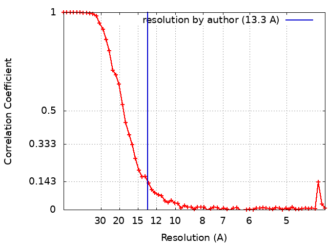
subtomogram averaging / cryo EM / Resolution: 13.3 Å
Sonderforschungsbereich 1035, project number 201302640, project B10
Germany
German Research Foundation (DFG)
392923329
Germany
Wellcome Trust
103139
United Kingdom
Wellcome Trust
203149
United Kingdom
Citation
Journal: Nat Commun / Year: 2021 Title: Structural basis for VPS34 kinase activation by Rab1 and Rab5 on membranes. Authors: Shirley Tremel / Yohei Ohashi / Dustin R Morado / Jessie Bertram / Olga Perisic / Laura T L Brandt / Marie-Kristin von Wrisberg / Zhuo A Chen / Sarah L Maslen / Oleksiy Kovtun / Mark Skehel ...Authors: Shirley Tremel / Yohei Ohashi / Dustin R Morado / Jessie Bertram / Olga Perisic / Laura T L Brandt / Marie-Kristin von Wrisberg / Zhuo A Chen / Sarah L Maslen / Oleksiy Kovtun / Mark Skehel / Juri Rappsilber / Kathrin Lang / Sean Munro / John A G Briggs / Roger L Williams / Abstract: The lipid phosphatidylinositol-3-phosphate (PI3P) is a regulator of two fundamental but distinct cellular processes, endocytosis and autophagy, so its generation needs to be under precise temporal ...The lipid phosphatidylinositol-3-phosphate (PI3P) is a regulator of two fundamental but distinct cellular processes, endocytosis and autophagy, so its generation needs to be under precise temporal and spatial control. PI3P is generated by two complexes that both contain the lipid kinase VPS34: complex II on endosomes (VPS34/VPS15/Beclin 1/UVRAG), and complex I on autophagosomes (VPS34/VPS15/Beclin 1/ATG14L). The endosomal GTPase Rab5 binds complex II, but the mechanism of VPS34 activation by Rab5 has remained elusive, and no GTPase is known to bind complex I. Here we show that Rab5a-GTP recruits endocytic complex II to membranes and activates it by binding between the VPS34 C2 and VPS15 WD40 domains. Electron cryotomography of complex II on Rab5a-decorated vesicles shows that the VPS34 kinase domain is released from inhibition by VPS15 and hovers over the lipid bilayer, poised for catalysis. We also show that the GTPase Rab1a, which is known to be involved in autophagy, recruits and activates the autophagy-specific complex I, but not complex II. Both Rabs bind to the same VPS34 interface but in a manner unique for each. These findings reveal how VPS34 complexes are activated on membranes by specific Rab GTPases and how they are recruited to unique cellular locations.
History
Deposition
Jan 23, 2021
-
Header (metadata) release
Jun 16, 2021
-
Map release
Jun 16, 2021
-
Update
Jun 16, 2021
-
Current status
Jun 16, 2021
Processing site: PDBe / Status: Released
-
Structure visualization
Movie
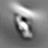
Surface view with section colored by density value
Download / File: emd_12237.map.gz / Format: CCP4 / Size: 125 MB / Type: IMAGE STORED AS FLOATING POINT NUMBER (4 BYTES)
Annotation
A reconstruction of the VPS34 complex II bound to Rab5a on lipid vesicles created by subtomogram averaging. The map was sharpened with LAFTER and resampled onto the map from EMD-12214
Voxel size
X=Y=Z: 1.0665 Å
Density
Contour Level
By AUTHOR: 0.008 / Movie #1: 0.008
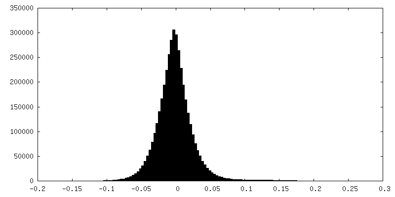
Minimum - Maximum
-0.08853249 - 0.12369968
Average (Standard dev.)
2.3871984e-05 (±0.005715127)
Symmetry
Space group: 1
Details
EMDB XML:
Map geometry
Axis order
X
Y
Z
Origin
0
0
0
Dimensions
320
320
320
Spacing
320
320
320
Cell
A=B=C: 341.27997 Å α=β=γ: 90.0 °
CCP4 map header:
mode
Image stored as Reals
Å/pix. X/Y/Z
1.0665
1.0665
1.0665
M x/y/z
320
320
320
origin x/y/z
0.000
0.000
0.000
length x/y/z
341.280
341.280
341.280
α/β/γ
90.000
90.000
90.000
start NX/NY/NZ
69
68
88
NX/NY/NZ
82
80
48
MAP C/R/S
1
2
3
start NC/NR/NS
0
0
0
NC/NR/NS
320
320
320
D min/max/mean
-0.089
0.124
0.000
-
Supplemental data
-
Half map: A reconstruction of the VPS34 complex II bound...
A reconstruction of the VPS34 complex II bound to Rab5a on lipid vesicles created by subtomogram averaging. The even half map, before LAFTER sharpening.
A reconstruction of the VPS34 complex II bound to Rab5a on lipid vesicles created by subtomogram averaging. The odd half map, before LAFTER sharpening.
Entire : human VPS34 complex II (VPS34-VPS15-Beclin1-UVRAG) bound to human...
Entire
Name: human VPS34 complex II (VPS34-VPS15-Beclin1-UVRAG) bound to human Rab5a on lipid vesicles
Components
Complex: human VPS34 complex II (VPS34-VPS15-Beclin1-UVRAG) bound to human Rab5a on lipid vesicles
Protein or peptide: UV radiation resistance-associated gene protein
Protein or peptide: Phosphatidylinositol 3-kinase catalytic subunit type 3
Protein or peptide: Phosphoinositide 3-kinase regulatory subunit 4
Protein or peptide: Beclin-1
Protein or peptide: Ras-related protein Rab-5A
-
Supramolecule #1: human VPS34 complex II (VPS34-VPS15-Beclin1-UVRAG) bound to human...
Supramolecule
Name: human VPS34 complex II (VPS34-VPS15-Beclin1-UVRAG) bound to human Rab5a on lipid vesicles type: complex / ID: 1 / Parent: 0 / Macromolecule list: all Details: The subunits were expressed using transient transfection in HEK293T cells. Cells were transfected with three plasmids: pYO1025 (encoding VPS34 and VPS15 in a pCAG backbone), pYO1124 ...Details: The subunits were expressed using transient transfection in HEK293T cells. Cells were transfected with three plasmids: pYO1025 (encoding VPS34 and VPS15 in a pCAG backbone), pYO1124 (encoding UVGRAG 1-464 fused to the BATS of ATG14, residues 413-492 in pVAG) and pYO1006 (Beclin1 in pCAG)
Source (natural)
Organism: Homo sapiens (human)
Recombinant expression
Organism: Homo sapiens (human)
Molecular weight
Theoretical: 392.16 KDa
-
Macromolecule #1: UV radiation resistance-associated gene protein
Model: Quantifoil / Material: GOLD / Mesh: 300 / Support film - Material: CARBON / Support film - topology: HOLEY ARRAY / Pretreatment - Type: GLOW DISCHARGE / Pretreatment - Atmosphere: AIR / Details: Quorum SC7620
Vitrification
Cryogen name: ETHANE / Chamber humidity: 100 % / Chamber temperature: 316 K / Instrument: FEI VITROBOT MARK I / Details: blot force was 20, with a blot time of 6 s.
-
Electron microscopy
Microscope
FEI TITAN KRIOS
Image recording
Film or detector model: GATAN K3 (6k x 4k) / Digitization - Dimensions - Width: 6000 pixel / Digitization - Dimensions - Height: 4000 pixel / Digitization - Sampling interval: 5.0 µm / Number grids imaged: 1 / Average exposure time: 0.55 sec. / Average electron dose: 2.99 e/Å2
Electron beam
Acceleration voltage: 300 kV / Electron source: FIELD EMISSION GUN
Number classes used: 6 / Applied symmetry - Point group: C1 (asymmetric) / Algorithm: FOURIER SPACE / Resolution.type: BY AUTHOR / Resolution: 13.3 Å / Resolution method: FSC 0.143 CUT-OFF / Software - Name: RELION (ver. 3.1) / Number subtomograms used: 26979
Extraction
Number tomograms: 105 / Number images used: 191196 Reference model: two gaussian-filtered elipsoids forming a V shape Details: To identify particles, subtomograms were aligned to an initial reference consisting of two gaussian-filtered ellipsoids forming a V-shape (Supplementary Fig. 6a, reference). After aligning ...Details: To identify particles, subtomograms were aligned to an initial reference consisting of two gaussian-filtered ellipsoids forming a V-shape (Supplementary Fig. 6a, reference). After aligning against the V-shape, some subtomograms converged and formed clusters, which indicated the presence of a particle The subtomogram coordinates were cleaned by a minimal distance threshold (distance cut off 8 px, cluster size 2, cluster distance 2 px) and cross correlation cut-off so that 191,169 particles remained.
The model in PDB 7BL1 was built and refined into the subtomogram averaged density map in EMD-12214. This map had the membrane portion of the reconstruction masked out. The reconstruction described in the final reconstruction described above includes the membrane that was masked out in EMD-12214. The final reconstruction was sharpened in LAFTER then resampled onto the density of EMD-12214, for ease of fitting the 7BL1 model.
Refinement
Space: REAL / Protocol: RIGID BODY FIT / Overall B value: 320 / Target criteria: Correlation coefficient
+
About Yorodumi
-
News
-
Feb 9, 2022. New format data for meta-information of EMDB entries
New format data for meta-information of EMDB entries
Version 3 of the EMDB header file is now the official format.
The previous official version 1.9 will be removed from the archive.
In the structure databanks used in Yorodumi, some data are registered as the other names, "COVID-19 virus" and "2019-nCoV". Here are the details of the virus and the list of structure data.
Jan 31, 2019. EMDB accession codes are about to change! (news from PDBe EMDB page)
EMDB accession codes are about to change! (news from PDBe EMDB page)
The allocation of 4 digits for EMDB accession codes will soon come to an end. Whilst these codes will remain in use, new EMDB accession codes will include an additional digit and will expand incrementally as the available range of codes is exhausted. The current 4-digit format prefixed with “EMD-” (i.e. EMD-XXXX) will advance to a 5-digit format (i.e. EMD-XXXXX), and so on. It is currently estimated that the 4-digit codes will be depleted around Spring 2019, at which point the 5-digit format will come into force.
The EM Navigator/Yorodumi systems omit the EMD- prefix.
Related info.:Q: What is EMD? / ID/Accession-code notation in Yorodumi/EM Navigator
Yorodumi is a browser for structure data from EMDB, PDB, SASBDB, etc.
This page is also the successor to EM Navigator detail page, and also detail information page/front-end page for Omokage search.
The word "yorodu" (or yorozu) is an old Japanese word meaning "ten thousand". "mi" (miru) is to see.
Related info.:EMDB / PDB / SASBDB / Comparison of 3 databanks / Yorodumi Search / Aug 31, 2016. New EM Navigator & Yorodumi / Yorodumi Papers / Jmol/JSmol / Function and homology information / Changes in new EM Navigator and Yorodumi
 Movie
Movie Controller

 Yorodumi
 Open data
 Basic information
 Map data
 Sample
 Function and homology information
 Homo sapiens (human)
 Authors
 United Kingdom,
  Germany, 9 items
  Citation
 Structure visualization
 Downloads & links
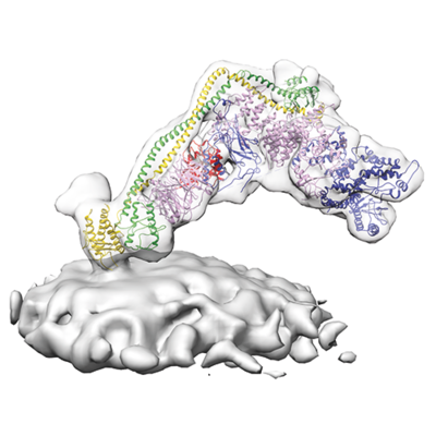 emd_12237.png
 http://ftp.pdbj.org/pub/emdb/structures/EMD-12237


 Z
Z Y
Y X
X


 Sample components
 Processing
 Electron microscopy
 FIELD EMISSION GUN