 Movie
Movie Controller


+ Open data

- Basic information
| Entry | Database: PDB / ID: 6ztz | |||||||||
|---|---|---|---|---|---|---|---|---|---|---|
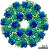
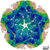
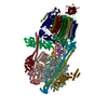
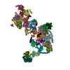
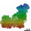
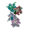
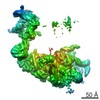
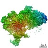
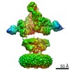
| Title | Assembly intermediates of orthoreovirus captured in the cell | |||||||||
 Components Components |
| |||||||||
 Keywords Keywords | VIRUS LIKE PARTICLE / Assemble Intermediates / Orthoreovirus / cryo-electron tomography / cellular lamellae | |||||||||
| Function / homology |  Function and homology information Function and homology informationicosahedral viral capsid / T=2 icosahedral viral capsid / host cell surface binding / viral inner capsid / symbiont-mediated suppression of host PKR/eIFalpha signaling / viral outer capsid / symbiont entry into host cell via permeabilization of host membrane / host cell endoplasmic reticulum / host cell mitochondrion / 7-methylguanosine mRNA capping ...icosahedral viral capsid / T=2 icosahedral viral capsid / host cell surface binding / viral inner capsid / symbiont-mediated suppression of host PKR/eIFalpha signaling / viral outer capsid / symbiont entry into host cell via permeabilization of host membrane / host cell endoplasmic reticulum / host cell mitochondrion / 7-methylguanosine mRNA capping / protein serine/threonine kinase inhibitor activity / viral life cycle / viral capsid / mRNA guanylyltransferase / mRNA guanylyltransferase activity / mRNA (guanine-N7)-methyltransferase / mRNA 5'-cap (guanine-N7-)-methyltransferase activity / RNA helicase activity / symbiont-mediated suppression of host innate immune response / RNA helicase / symbiont-mediated suppression of host type I interferon-mediated signaling pathway / GTP binding / host cell plasma membrane / structural molecule activity / ATP hydrolysis activity / RNA binding / zinc ion binding / ATP binding Similarity search - Function | |||||||||
| Biological species |  Reovirus sp. Reovirus sp. | |||||||||
| Method | ELECTRON MICROSCOPY / electron tomography / cryo EM / Resolution: 6.5 Å | |||||||||
 Authors Authors | Sutton, G.C. / Stuart, D.I. | |||||||||
| Funding support |  United Kingdom, 2items United Kingdom, 2items
| |||||||||
 Citation Citation | Journal: Nat Commun / Year: 2020 Title: Assembly intermediates of orthoreovirus captured in the cell. Authors: Geoff Sutton / Dapeng Sun / Xiaofeng Fu / Abhay Kotecha / Corey W Hecksel / Daniel K Clare / Peijun Zhang / David I Stuart / Mark Boyce /   Abstract: Traditionally, molecular assembly pathways for viruses are inferred from high resolution structures of purified stable intermediates, low resolution images of cell sections and genetic approaches. ...Traditionally, molecular assembly pathways for viruses are inferred from high resolution structures of purified stable intermediates, low resolution images of cell sections and genetic approaches. Here, we directly visualise an unsuspected 'single shelled' intermediate for a mammalian orthoreovirus in cryo-preserved infected cells, by cryo-electron tomography of cellular lamellae. Particle classification and averaging yields structures to 5.6 Å resolution, sufficient to identify secondary structural elements and produce an atomic model of the intermediate, comprising 120 copies each of protein λ1 and σ2. This λ1 shell is 'collapsed' compared to the mature virions, with molecules pushed inwards at the icosahedral fivefolds by ~100 Å, reminiscent of the first assembly intermediate of certain prokaryotic dsRNA viruses. This supports the supposition that these viruses share a common ancestor, and suggests mechanisms for the assembly of viruses of the Reoviridae. Such methodology holds promise for dissecting the replication cycle of many viruses. | |||||||||
| History |
|
- Structure visualization
| Movie |
Movie viewer |
|---|---|
| Structure viewer | Molecule: MolmilJmol/JSmol |
- Downloads & links
-Download
| PDBx/mmCIF format | 6ztz.cif.gz | 1.2 MB | Display | PDBx/mmCIF format |
|---|---|---|---|---|
| PDB format | pdb6ztz.ent.gz | 1012.9 KB | Display | PDB format |
| PDBx/mmJSON format | 6ztz.json.gz | Tree view | PDBx/mmJSON format | |
| Others |  Other downloads Other downloads |
-Validation report
| Arichive directory | https://data.pdbj.org/pub/pdb/validation_reports/zt/6ztzftp://data.pdbj.org/pub/pdb/validation_reports/zt/6ztz | HTTPS FTP |
|---|
-Related structure data
| Related structure data | 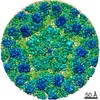 22166MC 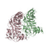 6xf7C 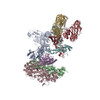 6xf8C  6ztsC 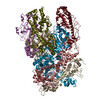 6ztyC M: map data used to model this data C: citing same article ( |
|---|---|
| Similar structure data |

-Links
 PDBj
PDBj


- Assembly
| Deposited unit | 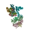
|
|---|---|
| 1 |
|
-Components
-Inner capsid protein lambda- ... , 2 types, 2 molecules BC
| #1: Protein | Mass: 116214.484 Da / Num. of mol.: 1 / Source method: isolated from a natural source / Source: (natural) Reovirus sp. / References: UniProt: Q9WAB2 |
|---|---|
| #2: Protein | Mass: 113187.195 Da / Num. of mol.: 1 / Source method: isolated from a natural source / Source: (natural) Reovirus sp. / References: UniProt: Q9WAB2 |
-Protein , 1 types, 2 molecules DP
| #3: Protein | Mass: 47024.016 Da / Num. of mol.: 2 / Source method: isolated from a natural source / Source: (natural) Reovirus sp. / References: UniProt: P11314 |
|---|
-Outer capsid protein ... , 3 types, 7 molecules KLMOXYZ
| #4: Protein | Mass: 69315.602 Da / Num. of mol.: 3 / Source method: isolated from a natural source / Source: (natural) Reovirus sp. / References: UniProt: P11077#5: Protein | | Mass: 143665.281 Da / Num. of mol.: 1 / Source method: isolated from a natural source / Source: (natural) Reovirus sp. / References: UniProt: P11079#6: Protein | Mass: 41237.117 Da / Num. of mol.: 3 / Source method: isolated from a natural source / Source: (natural) Reovirus sp. / References: UniProt: P07939 |
|---|
-Details
| Has protein modification | N |
|---|
-Experimental details
-Experiment
| Experiment | Method: ELECTRON MICROSCOPY |
|---|---|
| EM experiment | Aggregation state: CELL / 3D reconstruction method: electron tomography |
- Sample preparation
| Component | Name: Inner capsid protein lambda-1, Inner capsid protein sigma-2, Outer capsid protein mu-1, Outer capsid protein lambda-2 and Outer capsid protein sigma-3 Type: VIRUS / Entity ID: all / Source: NATURAL |
|---|---|
| Molecular weight | Experimental value: NO |
| Source (natural) | Organism: Reovirus sp. |
| Details of virus | Empty: YES / Enveloped: NO / Isolate: OTHER / Type: VIRION |
| Buffer solution | pH: 7 |
| Specimen | Embedding applied: NO / Shadowing applied: NO / Staining applied: NO / Vitrification applied: YES |
| Vitrification | Cryogen name: NITROGEN |
- Electron microscopy imaging
| Experimental equipment |  Model: Titan Krios / Image courtesy: FEI Company |
|---|---|
| Microscopy | Model: FEI TITAN KRIOS |
| Electron gun | Electron source:  FIELD EMISSION GUN / Accelerating voltage: 300 kV / Illumination mode: FLOOD BEAM FIELD EMISSION GUN / Accelerating voltage: 300 kV / Illumination mode: FLOOD BEAM |
| Electron lens | Mode: OTHER |
| Image recording | Electron dose: 2 e/Å2 / Film or detector model: GATAN K2 SUMMIT (4k x 4k) |
- Processing
| CTF correction | Type: PHASE FLIPPING AND AMPLITUDE CORRECTION |
|---|---|
| 3D reconstruction | Resolution: 6.5 Å / Resolution method: FSC 0.143 CUT-OFF |
| Atomic model building | Protocol: FLEXIBLE FIT |
