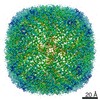
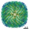
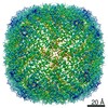
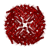
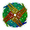
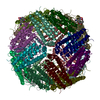
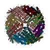
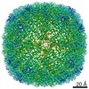
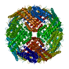
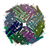
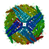
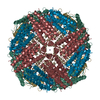
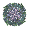
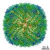
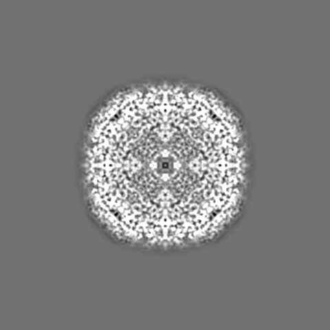
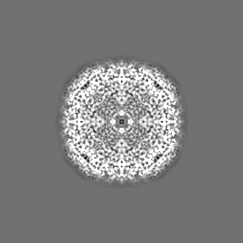
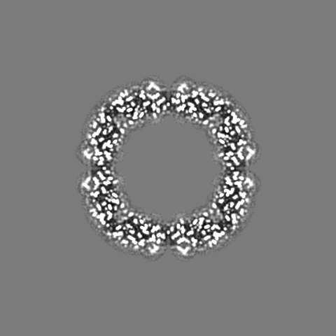
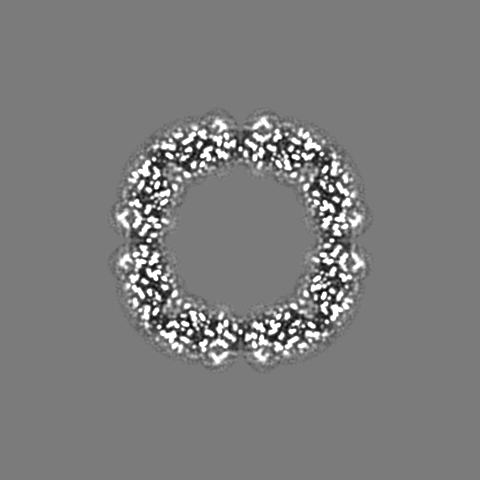
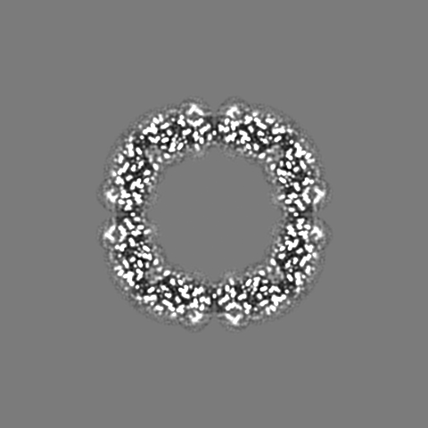
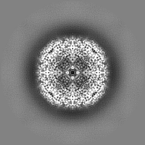
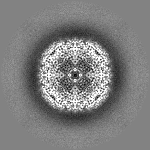
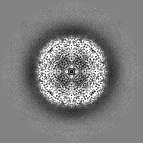
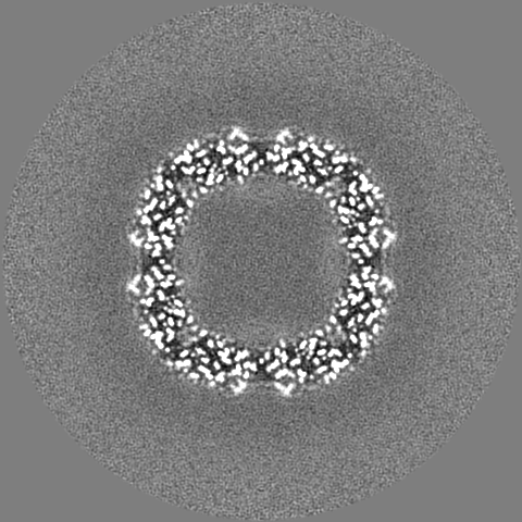
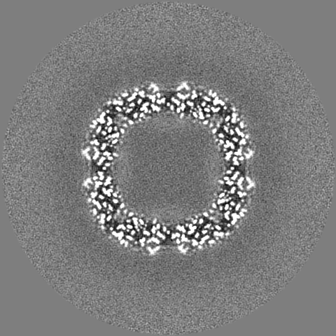
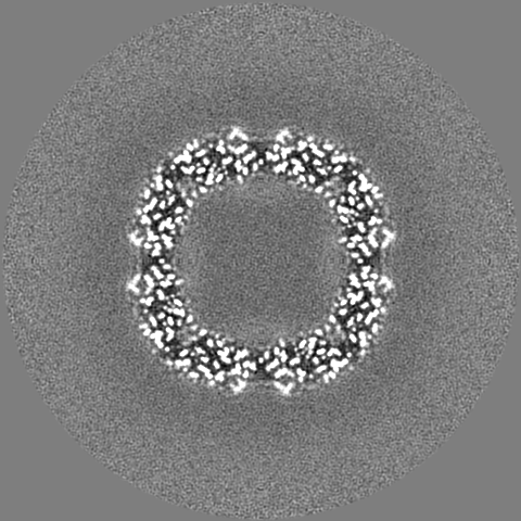
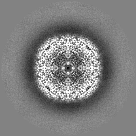
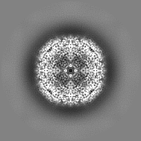
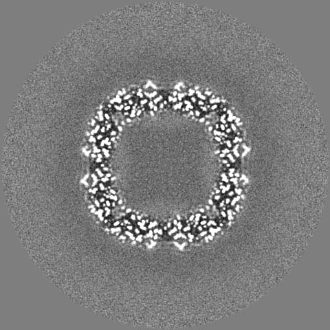
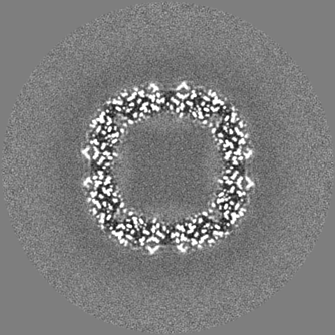
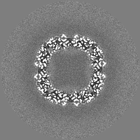
- EMDB-11122: 1.56 A structure of human apoferritin obtained from data subset o... -
+
データを開く
IDまたはキーワード:
読み込み中...
-
基本情報
登録情報
データベース: EMDB / ID: EMD-11122
タイトル
1.56 A structure of human apoferritin obtained from data subset of Titan Mono-BCOR microscope
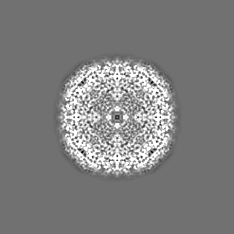
マップデータ
Sharpened map.
試料
細胞器官・細胞要素: Apoferritin
タンパク質・ペプチド: Ferritin heavy chain
リガンド: SODIUM ION
リガンド: water
機能・相同性
機能・相同性情報
iron ion sequestering activity / : / autolysosome / Scavenging by Class A Receptors / Golgi Associated Vesicle Biogenesis / ferroxidase / intracellular sequestering of iron ion / ferroxidase activity / negative regulation of fibroblast proliferation / ferric iron binding ...iron ion sequestering activity / : / autolysosome / Scavenging by Class A Receptors / Golgi Associated Vesicle Biogenesis / ferroxidase / intracellular sequestering of iron ion / ferroxidase activity / negative regulation of fibroblast proliferation / ferric iron binding / Iron uptake and transport / ferrous iron binding / tertiary granule lumen / iron ion transport / intracellular iron ion homeostasis / ficolin-1-rich granule lumen / iron ion binding / immune response / negative regulation of cell population proliferation / Neutrophil degranulation / extracellular exosome / extracellular region / identical protein binding / membrane / nucleus / cytoplasm / cytosol 類似検索 - 分子機能
ジャーナル: Nature / 年: 2020 タイトル: Atomic-resolution protein structure determination by cryo-EM. 著者: Ka Man Yip / Niels Fischer / Elham Paknia / Ashwin Chari / Holger Stark / 要旨: Single-particle electron cryo-microscopy (cryo-EM) is a powerful method for solving the three-dimensional structures of biological macromolecules. The technological development of transmission ...Single-particle electron cryo-microscopy (cryo-EM) is a powerful method for solving the three-dimensional structures of biological macromolecules. The technological development of transmission electron microscopes, detectors and automated procedures in combination with user-friendly image processing software and ever-increasing computational power have made cryo-EM a successful and expanding technology over the past decade. At resolutions better than 4 Å, atomic model building starts to become possible, but the direct visualization of true atomic positions in protein structure determination requires much higher (better than 1.5 Å) resolution, which so far has not been attained by cryo-EM. The direct visualization of atom positions is essential for understanding the mechanisms of protein-catalysed chemical reactions, and for studying how drugs bind to and interfere with the function of proteins. Here we report a 1.25 Å-resolution structure of apoferritin obtained by cryo-EM with a newly developed electron microscope that provides, to our knowledge, unprecedented structural detail. Our apoferritin structure has almost twice the 3D information content of the current world record reconstruction (at 1.54 Å resolution). We can visualize individual atoms in a protein, see density for hydrogen atoms and image single-atom chemical modifications. Beyond the nominal improvement in resolution, we also achieve a substantial improvement in the quality of the cryo-EM density map, which is highly relevant for using cryo-EM in structure-based drug design.
EMPIAR-10591 (タイトル: Atomic resolution structure of apoferritin from Titan Mono/BCorr microscope Data size: 41.7 TB Data #1: Single particle cryo-EM dataset of apoferritin from Titan Mono-BCorr microscope at 1.25 angstrom resolution [micrographs - multiframe])
 ムービー
ムービー コントローラー


 データを開く
 基本情報
基本情報 マップデータ
 試料
試料 機能・相同性情報
 Homo sapiens (ヒト)
 データ登録者
 ドイツ, 2件
  引用
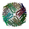
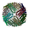
引用 構造の表示
構造の表示
 ダウンロードとリンク
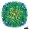
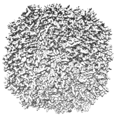 emd_11122.png
 http://ftp.pdbj.org/pub/emdb/structures/EMD-11122


 Z
Z Y
Y X
X

 試料の構成要素

 解析
解析 電子顕微鏡法
 FIELD EMISSION GUN
