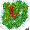
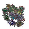
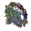
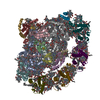
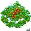
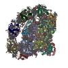
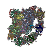
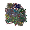
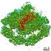
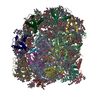
Complex: Triple complex of photosystem I bound to ferredoxin and plastocyanin
Protein or peptide: x 14 types
Protein or peptide: x 4 types
Ligand: x 18 types
Keywords
Membrane complex / photosystem I / ferredoxin / light harvesting / excitation transfer / PHOTOSYNTHESIS
Function / homology
Function and homology information
: / photosynthesis, light harvesting / chloroplast thylakoid lumen / photosystem I reaction center / photosystem I / photosynthetic electron transport in photosystem I / photosystem I / photosystem II / chloroplast stroma / chlorophyll binding ...: / photosynthesis, light harvesting / chloroplast thylakoid lumen / photosystem I reaction center / photosystem I / photosynthetic electron transport in photosystem I / photosystem I / photosystem II / chloroplast stroma / chlorophyll binding / chloroplast thylakoid membrane / photosynthesis / chloroplast / electron transport chain / 2 iron, 2 sulfur cluster binding / 4 iron, 4 sulfur cluster binding / electron transfer activity / oxidoreductase activity / copper ion binding / magnesium ion binding / metal ion binding Similarity search - Function
Photosystem I PsaH, reaction centre subunit VI / Photosystem I reaction centre subunit VI / Plastocyanin / 4Fe-4S dicluster domain / Photosystem I reaction center subunit psaK, plant / Photosystem I reaction center subunit V/PsaK, plant / Photosystem I PsaG/PsaK domain, chloroplastic / Ferredoxin [2Fe-2S], plant / Photosystem I reaction centre subunit PsaK superfamily / Blue (type 1) copper protein, plastocyanin-type ...Photosystem I PsaH, reaction centre subunit VI / Photosystem I reaction centre subunit VI / Plastocyanin / 4Fe-4S dicluster domain / Photosystem I reaction center subunit psaK, plant / Photosystem I reaction center subunit V/PsaK, plant / Photosystem I PsaG/PsaK domain, chloroplastic / Ferredoxin [2Fe-2S], plant / Photosystem I reaction centre subunit PsaK superfamily / Blue (type 1) copper protein, plastocyanin-type / Photosystem I reaction center subunit V/PsaK / Photosystem I psaG / psaK / Photosystem I PsaL, reaction centre subunit XI / Photosystem I, reaction centre subunit XI / Photosystem I PsaL, reaction centre subunit XI superfamily / Photosystem I reaction centre subunit XI / Photosystem I reaction centre subunit VIII / Photosystem I reaction centre subunit VIII / Photosystem I reaction centre subunit VIII superfamily / Photosystem I PsaF, reaction centre subunit III / Photosystem I PsaF, reaction centre subunit III superfamily / Photosystem I reaction centre subunit III / Photosystem I PsaD / Photosystem I, reaction centre subunit PsaD superfamily / PsaD / Photosystem I PsaE, reaction centre subunit IV / Photosystem I PsaJ, reaction centre subunit IX superfamily / Photosystem I reaction centre subunit IV / PsaE / Chlorophyll A-B binding protein, plant and chromista / Photosystem I PsaJ, reaction centre subunit IX / Photosystem I reaction centre subunit IX / PsaJ / Chlorophyll A-B binding protein / Chlorophyll A-B binding protein / Photosystem I protein PsaC / Photosystem I PsaB / Photosystem I PsaA / Photosystem I PsaA/PsaB, conserved site / Photosystem I psaA and psaB proteins signature. / : / Photosystem I PsaA/PsaB / Photosystem I PsaA/PsaB superfamily / Photosystem I psaA/psaB protein / 2Fe-2S ferredoxin, iron-sulphur binding site / 2Fe-2S ferredoxin-type iron-sulfur binding region signature. / Blue (type 1) copper domain / Copper binding proteins, plastocyanin/azurin family / Blue (type 1) copper protein, binding site / Type-1 copper (blue) proteins signature. / Electron transport accessory-like domain superfamily / 2Fe-2S iron-sulfur cluster binding domain / Beta-grasp domain superfamily / 2Fe-2S ferredoxin-type iron-sulfur binding domain profile. / 2Fe-2S ferredoxin-type iron-sulfur binding domain / 2Fe-2S ferredoxin-like superfamily / 4Fe-4S ferredoxin, iron-sulphur binding, conserved site / 4Fe-4S ferredoxin-type iron-sulfur binding region signature. / 4Fe-4S ferredoxin-type iron-sulfur binding domain profile. / 4Fe-4S ferredoxin-type, iron-sulphur binding domain / Cupredoxin Similarity search - Domain/homology
Photosystem I P700 chlorophyll a apoprotein A1 / Photosystem I P700 chlorophyll a apoprotein A2 / Photosystem I reaction center subunit VI / Photosystem I reaction center subunit III / Photosystem I reaction center subunit IX / Photosystem I reaction center subunit IV / Photosystem I reaction center subunit II, chloroplastic / Photosystem I reaction center subunit XI, chloroplastic / Photosystem I reaction center subunit psaK, chloroplastic / Ferredoxin-1, chloroplastic ...Photosystem I P700 chlorophyll a apoprotein A1 / Photosystem I P700 chlorophyll a apoprotein A2 / Photosystem I reaction center subunit VI / Photosystem I reaction center subunit III / Photosystem I reaction center subunit IX / Photosystem I reaction center subunit IV / Photosystem I reaction center subunit II, chloroplastic / Photosystem I reaction center subunit XI, chloroplastic / Photosystem I reaction center subunit psaK, chloroplastic / Ferredoxin-1, chloroplastic / Photosystem I iron-sulfur center / Plastocyanin, chloroplastic / Photosystem I reaction center subunit VIII / Chlorophyll a-b binding protein 3, chloroplastic / Chlorophyll a-b binding protein, chloroplastic / Chlorophyll a-b binding protein P4, chloroplastic Similarity search - Component
Biological species
Pisum sativum (garden pea)
Method
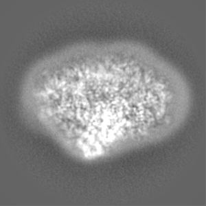
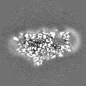
single particle reconstruction / cryo EM / Resolution: 2.7 Å
German-Israeli Foundation for Research and Development
1483
Israel
Citation
Journal: Nat Plants / Year: 2020 Title: The structure of a triple complex of plant photosystem I with ferredoxin and plastocyanin. Authors: Ido Caspy / Anna Borovikova-Sheinker / Daniel Klaiman / Yoel Shkolnisky / Nathan Nelson / Abstract: The ability of photosynthetic organisms to use sunlight as a sole source of energy is endowed by two large membrane complexes-photosystem I (PSI) and photosystem II (PSII). PSI and PSII are the ...The ability of photosynthetic organisms to use sunlight as a sole source of energy is endowed by two large membrane complexes-photosystem I (PSI) and photosystem II (PSII). PSI and PSII are the fundamental components of oxygenic photosynthesis, providing oxygen, food and an energy source for most living organisms on Earth. Currently, high-resolution crystal structures of these complexes from various organisms are available. The crystal structures of megadalton complexes have revealed excitation transfer and electron-transport pathways within the various complexes. PSI is defined as plastocyanin-ferredoxin oxidoreductase but a high-resolution structure of the entire triple supercomplex is not available. Here, using a new cryo-electron microscopy technique, we solve the structure of native plant PSI in complex with its electron donor plastocyanin and the electron acceptor ferredoxin. We reveal all of the contact sites and the modes of interaction between the interacting electron carriers and PSI.
History
Deposition
Mar 25, 2020
-
Header (metadata) release
Sep 30, 2020
-
Map release
Sep 30, 2020
-
Update
Oct 9, 2024
-
Current status
Oct 9, 2024
Processing site: PDBe / Status: Released
-
Structure visualization
Movie
Surface view with section colored by density value
Film or detector model: GATAN K3 BIOQUANTUM (6k x 4k) / Number real images: 11622 / Average exposure time: 1.4 sec. / Average electron dose: 47.05 e/Å2
Electron beam
Acceleration voltage: 300 kV / Electron source: FIELD EMISSION GUN
In the structure databanks used in Yorodumi, some data are registered as the other names, "COVID-19 virus" and "2019-nCoV". Here are the details of the virus and the list of structure data.
Jan 31, 2019. EMDB accession codes are about to change! (news from PDBe EMDB page)
EMDB accession codes are about to change! (news from PDBe EMDB page)
The allocation of 4 digits for EMDB accession codes will soon come to an end. Whilst these codes will remain in use, new EMDB accession codes will include an additional digit and will expand incrementally as the available range of codes is exhausted. The current 4-digit format prefixed with “EMD-” (i.e. EMD-XXXX) will advance to a 5-digit format (i.e. EMD-XXXXX), and so on. It is currently estimated that the 4-digit codes will be depleted around Spring 2019, at which point the 5-digit format will come into force.
The EM Navigator/Yorodumi systems omit the EMD- prefix.
Related info.:Q: What is EMD? / ID/Accession-code notation in Yorodumi/EM Navigator
Yorodumi is a browser for structure data from EMDB, PDB, SASBDB, etc.
This page is also the successor to EM Navigator detail page, and also detail information page/front-end page for Omokage search.
The word "yorodu" (or yorozu) is an old Japanese word meaning "ten thousand". "mi" (miru) is to see.
Related info.:EMDB / PDB / SASBDB / Comparison of 3 databanks / Yorodumi Search / Aug 31, 2016. New EM Navigator & Yorodumi / Yorodumi Papers / Jmol/JSmol / Function and homology information / Changes in new EM Navigator and Yorodumi
 Movie
Movie Controller


 Open data
 Basic information
 Map data
 Sample
 Keywords
 Function and homology information
 Pisum sativum (garden pea)
 Authors
 Israel, 2 items
  Citation
 Structure visualization
 Downloads & links
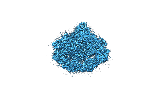 emd_10798.png
 http://ftp.pdbj.org/pub/emdb/structures/EMD-10798


 Z (Sec.)
 Y (Row.)
 X (Col.)


 Sample components


 Processing
 Electron microscopy
 FIELD EMISSION GUN


