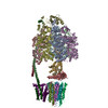
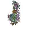
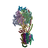
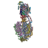
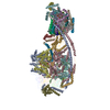
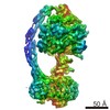
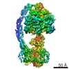
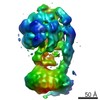
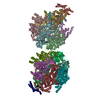
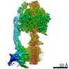
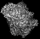
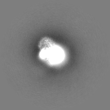
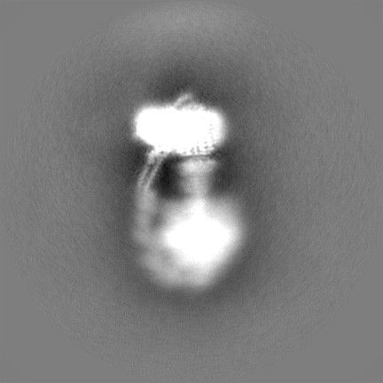
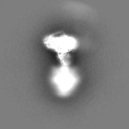
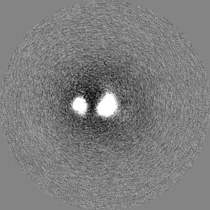
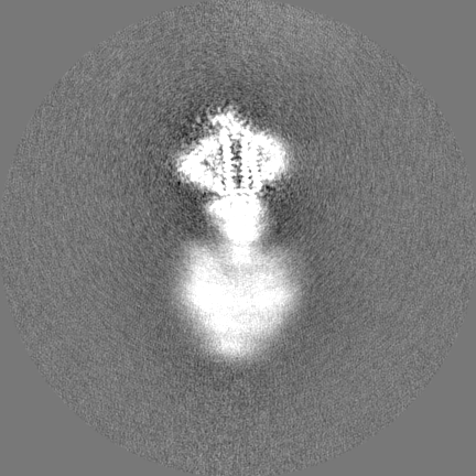
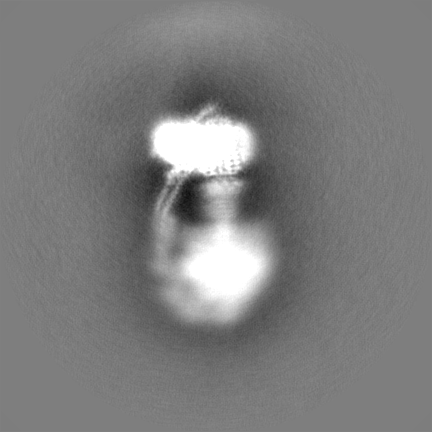
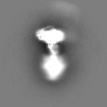
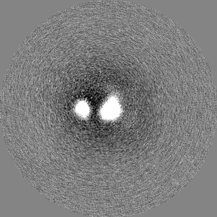
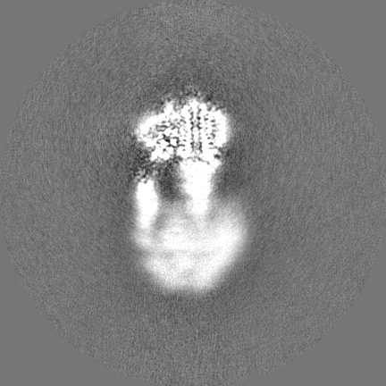
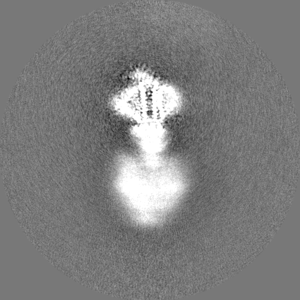
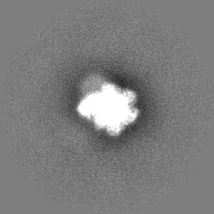
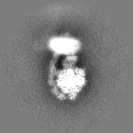
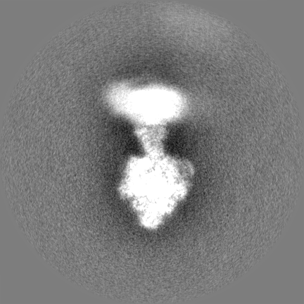
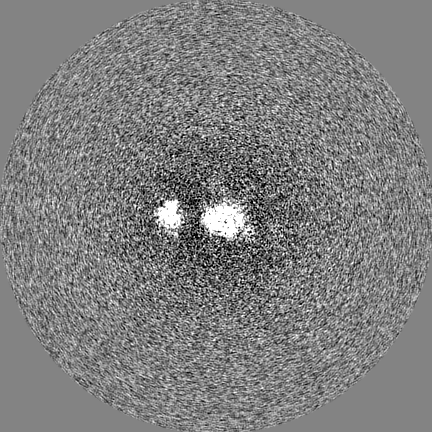
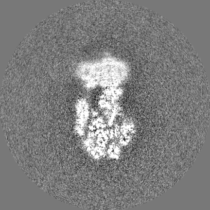
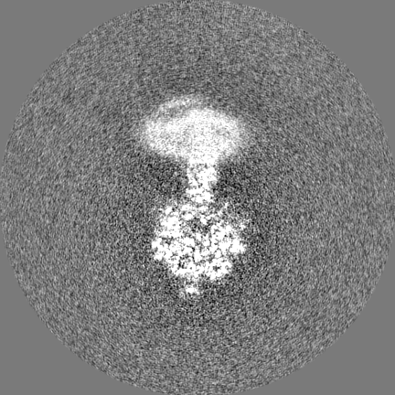
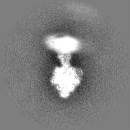
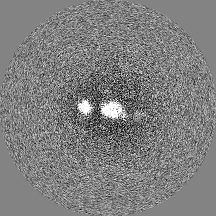
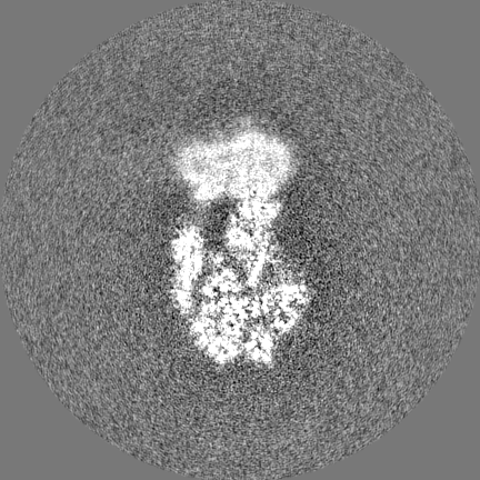
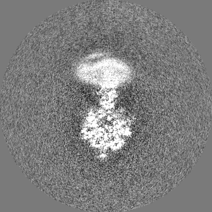
Composite map of state 1a of ovine F1Fo, obtained from series of focused refinements as described in the paper.
Sample
Complex: Purified ATP synthase from sheep heart mitochondria
Protein or peptide: x 17 types
Ligand: x 6 types
Keywords
ATP synthase / mitochondrial / respiratory chain / mammalian / MEMBRANE PROTEIN
Function / homology
Function and homology information
: / : / negative regulation of cell adhesion involved in substrate-bound cell migration / : / angiostatin binding / Formation of ATP by chemiosmotic coupling / Cristae formation / mitochondrial proton-transporting ATP synthase complex assembly / cellular response to interleukin-7 / proton channel activity ...: / : / negative regulation of cell adhesion involved in substrate-bound cell migration / : / angiostatin binding / Formation of ATP by chemiosmotic coupling / Cristae formation / mitochondrial proton-transporting ATP synthase complex assembly / cellular response to interleukin-7 / proton channel activity / Mitochondrial protein degradation / MHC class I protein binding / proton transmembrane transporter activity / proton motive force-driven ATP synthesis / proton-transporting two-sector ATPase complex, proton-transporting domain / mitochondrial nucleoid / proton motive force-driven mitochondrial ATP synthesis / proton-transporting ATPase activity, rotational mechanism / positive regulation of blood vessel endothelial cell migration / H+-transporting two-sector ATPase / proton-transporting ATP synthase complex / proton-transporting ATP synthase activity, rotational mechanism / proton transmembrane transport / aerobic respiration / regulation of intracellular pH / mitochondrial membrane / lipid metabolic process / angiogenesis / mitochondrial inner membrane / lipid binding / cell surface / mitochondrion / ATP binding / plasma membrane Similarity search - Function
ATP synthase membrane subunit K / ATP synthase regulation / ATP synthase subunit ATP5MJ, mitochondrial / Mitochondrial proteolipid / ATP synthase, F0 complex, subunit G, mitochondrial / ATP synthase, F0 complex, subunit E, mitochondrial / Mitochondrial ATP synthase subunit g, animal / Mitochondrial ATP synthase g subunit / ATP synthase E chain / ATP synthase protein 8, metazoa ...ATP synthase membrane subunit K / ATP synthase regulation / ATP synthase subunit ATP5MJ, mitochondrial / Mitochondrial proteolipid / ATP synthase, F0 complex, subunit G, mitochondrial / ATP synthase, F0 complex, subunit E, mitochondrial / Mitochondrial ATP synthase subunit g, animal / Mitochondrial ATP synthase g subunit / ATP synthase E chain / ATP synthase protein 8, metazoa / Mitochondrial F1-F0 ATP synthase subunit F, predicted / ATP synthase protein 8, mammals / ATP synthase protein 8 / Mitochondrial F1F0-ATP synthase, subunit f / ATP synthase-coupling factor 6, mitochondrial / ATP synthase-coupling factor 6 superfamily, mitochondrial / Mitochondrial ATP synthase coupling factor 6 / ATP synthase, F0 complex, subunit B/MI25 / ATP synthase, F0 complex, subunit B / Mitochondrial ATP synthase B chain precursor (ATP-synt_B) / ATP synthase delta/epsilon subunit, C-terminal domain superfamily / ATP synthase, F0 complex, subunit D, mitochondrial / ATP synthase D chain, mitochondrial (ATP5H) / ATP synthase, F0 complex, subunit D superfamily, mitochondrial / ATP synthase, F0 complex, subunit A, bacterial/mitochondria / ATP synthase, F1 complex, epsilon subunit, mitochondrial / ATP synthase, F1 complex, epsilon subunit superfamily, mitochondrial / Mitochondrial ATP synthase epsilon chain / ATPase, OSCP/delta subunit, conserved site / ATP synthase delta (OSCP) subunit signature. / F1F0 ATP synthase OSCP/delta subunit, N-terminal domain superfamily / ATP synthase, F0 complex, subunit A / ATP synthase, F0 complex, subunit A, active site / ATP synthase, F0 complex, subunit A superfamily / ATP synthase A chain / ATP synthase a subunit signature. / ATPase, OSCP/delta subunit / ATP synthase delta (OSCP) subunit / ATP synthase, F1 complex, delta/epsilon subunit / ATP synthase, F1 complex, delta/epsilon subunit, N-terminal / F0F1 ATP synthase delta/epsilon subunit, N-terminal / ATP synthase, Delta/Epsilon chain, beta-sandwich domain / ATP synthase, F0 complex, subunit C / F1F0 ATP synthase subunit C superfamily / ATP synthase, F0 complex, subunit C, DCCD-binding site / ATP synthase c subunit signature. / ATP synthase, F1 complex, gamma subunit conserved site / ATP synthase gamma subunit signature. / ATP synthase, F1 complex, beta subunit / ATP synthase, alpha subunit, C-terminal domain superfamily / ATP synthase, F1 complex, gamma subunit / ATP synthase, F1 complex, gamma subunit superfamily / ATP synthase / ATP synthase, F1 complex, alpha subunit nucleotide-binding domain / ATP synthase, alpha subunit, C-terminal / ATP synthase, F1 complex, alpha subunit / ATP synthase alpha/beta chain, C terminal domain / V-ATPase proteolipid subunit C-like domain / F/V-ATP synthase subunit C superfamily / ATP synthase subunit C / ATPase, F1/V1 complex, beta/alpha subunit, C-terminal / ATP synthase subunit alpha, N-terminal domain-like superfamily / ATPase, F1/V1/A1 complex, alpha/beta subunit, N-terminal domain superfamily / ATPase, F1/V1/A1 complex, alpha/beta subunit, N-terminal domain / ATP synthase alpha/beta family, beta-barrel domain / ATPase, alpha/beta subunit, nucleotide-binding domain, active site / ATP synthase alpha and beta subunits signature. / ATPase, F1/V1/A1 complex, alpha/beta subunit, nucleotide-binding domain / ATP synthase alpha/beta family, nucleotide-binding domain / ATPases associated with a variety of cellular activities / AAA+ ATPase domain / P-loop containing nucleoside triphosphate hydrolase Similarity search - Domain/homology
ATP synthase peripheral stalk subunit OSCP / ATP synthase F(0) complex subunit 8 / ATP synthase F(0) complex subunit a / ATP synthase F(1) complex subunit gamma, mitochondrial / ATP synthase F(0) complex subunit C1, mitochondrial / ATP synthase subunit alpha / Uncharacterized protein / Uncharacterized protein / ATP synthase subunit beta / ATP synthase F(0) complex subunit e, mitochondrial ...ATP synthase peripheral stalk subunit OSCP / ATP synthase F(0) complex subunit 8 / ATP synthase F(0) complex subunit a / ATP synthase F(1) complex subunit gamma, mitochondrial / ATP synthase F(0) complex subunit C1, mitochondrial / ATP synthase subunit alpha / Uncharacterized protein / Uncharacterized protein / ATP synthase subunit beta / ATP synthase F(0) complex subunit e, mitochondrial / ATP synthase F1 subunit delta / ATP synthase peripheral stalk subunit d, mitochondrial / ATP synthase peripheral stalk subunit OSCP, mitochondrial / ATP synthase peripheral stalk subunit F6, mitochondrial / ATP synthase subunit epsilon, mitochondrial / ATP synthase F(0) complex subunit g, mitochondrial / ATP synthase F(0) complex subunit f, mitochondrial / ATP synthase subunit b Similarity search - Component
Biological species
Ovis aries (sheep)
Method
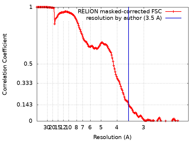
single particle reconstruction / cryo EM / Resolution: 3.5 Å
Journal: Nat Struct Mol Biol / Year: 2020 Title: Cryo-EM structure of the entire mammalian F-type ATP synthase. Authors: Gergely Pinke / Long Zhou / Leonid A Sazanov / Abstract: The majority of adenosine triphosphate (ATP) powering cellular processes in eukaryotes is produced by the mitochondrial F1Fo ATP synthase. Here, we present the atomic models of the membrane Fo domain ...The majority of adenosine triphosphate (ATP) powering cellular processes in eukaryotes is produced by the mitochondrial F1Fo ATP synthase. Here, we present the atomic models of the membrane Fo domain and the entire mammalian (ovine) F1Fo, determined by cryo-electron microscopy. Subunits in the membrane domain are arranged in the 'proton translocation cluster' attached to the c-ring and a more distant 'hook apparatus' holding subunit e. Unexpectedly, this subunit is anchored to a lipid 'plug' capping the c-ring. We present a detailed proton translocation pathway in mammalian Fo and key inter-monomer contacts in F1Fo multimers. Cryo-EM maps of F1Fo exposed to calcium reveal a retracted subunit e and a disassembled c-ring, suggesting permeability transition pore opening. We propose a model for the permeability transition pore opening, whereby subunit e pulls the lipid plug out of the c-ring. Our structure will allow the design of drugs for many emerging applications in medicine.
History
Deposition
Dec 23, 2019
-
Header (metadata) release
Sep 23, 2020
-
Map release
Sep 23, 2020
-
Update
May 22, 2024
-
Current status
May 22, 2024
Processing site: PDBe / Status: Released
-
Structure visualization
Movie
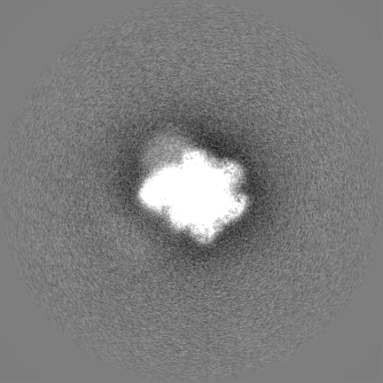
Surface view with section colored by density value
Cryogen name: ETHANE-PROPANE / Chamber humidity: 100 % / Chamber temperature: 277 K / Instrument: FEI VITROBOT MARK IV
-
Electron microscopy
Microscope
FEI TITAN KRIOS
Image recording
Film or detector model: FEI FALCON III (4k x 4k) / Detector mode: INTEGRATING / Digitization - Dimensions - Width: 4096 pixel / Digitization - Dimensions - Height: 4096 pixel / Number grids imaged: 1 / Number real images: 3053 / Average exposure time: 1.0 sec. / Average electron dose: 106.0 e/Å2 / Details: 40 frames per movie
Electron beam
Acceleration voltage: 300 kV / Electron source: FIELD EMISSION GUN
In the structure databanks used in Yorodumi, some data are registered as the other names, "COVID-19 virus" and "2019-nCoV". Here are the details of the virus and the list of structure data.
Jan 31, 2019. EMDB accession codes are about to change! (news from PDBe EMDB page)
EMDB accession codes are about to change! (news from PDBe EMDB page)
The allocation of 4 digits for EMDB accession codes will soon come to an end. Whilst these codes will remain in use, new EMDB accession codes will include an additional digit and will expand incrementally as the available range of codes is exhausted. The current 4-digit format prefixed with “EMD-” (i.e. EMD-XXXX) will advance to a 5-digit format (i.e. EMD-XXXXX), and so on. It is currently estimated that the 4-digit codes will be depleted around Spring 2019, at which point the 5-digit format will come into force.
The EM Navigator/Yorodumi systems omit the EMD- prefix.
Related info.:Q: What is EMD? / ID/Accession-code notation in Yorodumi/EM Navigator
Yorodumi is a browser for structure data from EMDB, PDB, SASBDB, etc.
This page is also the successor to EM Navigator detail page, and also detail information page/front-end page for Omokage search.
The word "yorodu" (or yorozu) is an old Japanese word meaning "ten thousand". "mi" (miru) is to see.
Related info.:EMDB / PDB / SASBDB / Comparison of 3 databanks / Yorodumi Search / Aug 31, 2016. New EM Navigator & Yorodumi / Yorodumi Papers / Jmol/JSmol / Function and homology information / Changes in new EM Navigator and Yorodumi
 Movie
Movie Controller


 Open data
 Basic information
 Map data
 Sample
 Keywords
 Function and homology information
 Authors
 Citation
 Structure visualization
 Downloads & links
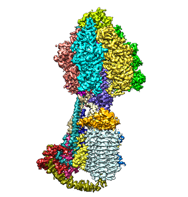 emd_10573.png
 http://ftp.pdbj.org/pub/emdb/structures/EMD-10573


 X (Sec.)
 Y (Row.)
 Z (Col.)


 Sample components


 Processing
 Electron microscopy
 FIELD EMISSION GUN