 Movie
Movie Controller


+ Open data

- Basic information
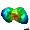
| Entry | Database: EMDB / ID: EMD-3098 | |||||||||
|---|---|---|---|---|---|---|---|---|---|---|
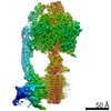
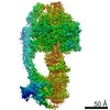
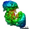
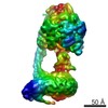
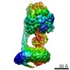
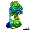
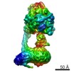
| Title | Cryo-EM structure of bovine FoF1 ATP synthase | |||||||||
 Map data Map data | 3D map of bovine FoF1 ATP synthase | |||||||||
 Sample Sample |
| |||||||||
 Keywords Keywords | mitochondria / bovine heart / Bos taurus / OXPHOS | |||||||||
| Biological species |  | |||||||||
| Method | single particle reconstruction / cryo EM / Resolution: 11.0 Å | |||||||||
 Authors Authors | Hauer F / Gerle C / Fischer N / Oshima A / Shinzawa-Itoh K / Shimada S / Yokoyama K / Fujiyoshi Y / Stark H | |||||||||
 Citation Citation | Journal: Structure / Year: 2015 Title: GraDeR: Membrane Protein Complex Preparation for Single-Particle Cryo-EM. Authors: Florian Hauer / Christoph Gerle / Niels Fischer / Atsunori Oshima / Kyoko Shinzawa-Itoh / Satoru Shimada / Ken Yokoyama / Yoshinori Fujiyoshi / Holger Stark /   Abstract: We developed a method, named GraDeR, which substantially improves the preparation of membrane protein complexes for structure determination by single-particle cryo-electron microscopy (cryo-EM). In ...We developed a method, named GraDeR, which substantially improves the preparation of membrane protein complexes for structure determination by single-particle cryo-electron microscopy (cryo-EM). In GraDeR, glycerol gradient centrifugation is used for the mild removal of free detergent monomers and micelles from lauryl maltose-neopentyl glycol detergent stabilized membrane complexes, resulting in monodisperse and stable complexes to which standard processes for water-soluble complexes can be applied. We demonstrate the applicability of the method on three different membrane complexes, including the mammalian FoF1 ATP synthase. For this highly dynamic and fragile rotary motor, we show that GraDeR allows visualizing the asymmetry of the F1 domain, which matches the ground state structure of the isolated domain. Therefore, the present cryo-EM structure of FoF1 ATP synthase provides direct structural evidence for Boyer's binding change mechanism in the context of the intact enzyme. | |||||||||
| History |
|
- Structure visualization
| Movie |
 Movie viewer Movie viewer |
|---|---|
| Structure viewer | EM map: SurfViewMolmilJmol/JSmol |
| Supplemental images |

- Downloads & links
-EMDB archive
| Map data | emd_3098.map.gz | 37.2 MB | EMDB map data format | |
|---|---|---|---|---|
| Header (meta data) | emd-3098-v30.xmlemd-3098.xml | 9.4 KB 9.4 KB | Display Display | EMDB header |
| Images | 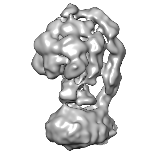 EMDB-3098.png EMDB-3098.png | 86.6 KB | ||
| Archive directory |  http://ftp.pdbj.org/pub/emdb/structures/EMD-3098ftp://ftp.pdbj.org/pub/emdb/structures/EMD-3098 http://ftp.pdbj.org/pub/emdb/structures/EMD-3098ftp://ftp.pdbj.org/pub/emdb/structures/EMD-3098 | HTTPS FTP |
-Related structure data
| Similar structure data |
|---|
-Links
| EMDB pages | EMDB (EBI/PDBe) / EMDataResource |
|---|
-Map
| File | Download / File: emd_3098.map.gz / Format: CCP4 / Size: 39.7 MB / Type: IMAGE STORED AS FLOATING POINT NUMBER (4 BYTES) | ||||||||||||||||||||||||||||||||||||||||||||||||||||||||||||||||||||
|---|---|---|---|---|---|---|---|---|---|---|---|---|---|---|---|---|---|---|---|---|---|---|---|---|---|---|---|---|---|---|---|---|---|---|---|---|---|---|---|---|---|---|---|---|---|---|---|---|---|---|---|---|---|---|---|---|---|---|---|---|---|---|---|---|---|---|---|---|---|
| Annotation | 3D map of bovine FoF1 ATP synthase | ||||||||||||||||||||||||||||||||||||||||||||||||||||||||||||||||||||
| Projections & slices | Image control
Images are generated by Spider. | ||||||||||||||||||||||||||||||||||||||||||||||||||||||||||||||||||||
| Voxel size | X=Y=Z: 1.6 Å | ||||||||||||||||||||||||||||||||||||||||||||||||||||||||||||||||||||
| Density |
| ||||||||||||||||||||||||||||||||||||||||||||||||||||||||||||||||||||
| Symmetry | Space group: 1 | ||||||||||||||||||||||||||||||||||||||||||||||||||||||||||||||||||||
| Details | EMDB XML:
CCP4 map header:
| ||||||||||||||||||||||||||||||||||||||||||||||||||||||||||||||||||||
 Z (Sec.)
 Y (Row.)
 X (Col.)


-Supplemental data
- Sample components
-Entire : Bovine mitochondrial FoF1 ATP synthase
| Entire | Name: Bovine mitochondrial FoF1 ATP synthase |
|---|---|
| Components |
|
-Supramolecule #1000: Bovine mitochondrial FoF1 ATP synthase
| Supramolecule | Name: Bovine mitochondrial FoF1 ATP synthase / type: sample / ID: 1000 Details: The sample was initially maintained in a buffer containing the detergent lauryl maltose-neopentyl glycol (LMNG). For cryo-EM preparation, free LMNG-monomers and micelles were removed using the GraDeR method. Oligomeric state: Monomer / Number unique components: 1 |
|---|---|
| Molecular weight | Experimental: 600 KDa / Theoretical: 600 KDa |
-Macromolecule #1: FoF1 ATP synthase
| Macromolecule | Name: FoF1 ATP synthase / type: protein_or_peptide / ID: 1 / Name.synonym: F-ATPase, ATP-synthase / Number of copies: 1 / Oligomeric state: Monomer / Recombinant expression: No |
|---|---|
| Source (natural) | Organism: |
| Molecular weight | Experimental: 600 KDa / Theoretical: 600 KDa |
-Experimental details
-Structure determination
| Method | cryo EM |
|---|---|
 Processing Processing | single particle reconstruction |
| Aggregation state | particle |
-Sample preparation
| Buffer | pH: 8 Details: 50 mM HEPES pH 8.0, 100 mM NaCl, 0.1% azide, 0.5 mM ADP and 5 mM MgCl2 |
|---|---|
| Grid | Details: 200 mesh copper Quantifoil grids (3.5/3.5) with custom-made holey carbon film covered by custom-made thin continuous carbon film |
| Vitrification | Cryogen name: ETHANE / Chamber humidity: 95 % / Instrument: FEI VITROBOT MARK IV Details: Nitrocellulose paper was used to allow for slow blotting to avoid disintegration of the FoF1 ATP synthase complex Method: Slow blotting (15 seconds) at low blot force |
- Electron microscopy
| Microscope | FEI TITAN KRIOS |
|---|---|
| Alignment procedure | Legacy - Astigmatism: Using a Cs-corrector from CEOS electron optical aberrations were corrected to residual phase errors of 45degree at scattering angles of >12 to 15 mrad |
| Date | Aug 20, 2013 |
| Image recording | Category: CCD / Film or detector model: FEI FALCON II (4k x 4k) / Number real images: 3956 / Average electron dose: 50 e/Å2 |
| Electron beam | Acceleration voltage: 300 kV / Electron source:  FIELD EMISSION GUN FIELD EMISSION GUN |
| Electron optics | Illumination mode: SPOT SCAN / Imaging mode: BRIGHT FIELD / Cs: 0.01 mm / Nominal defocus max: 5.0 µm / Nominal defocus min: 2.5 µm / Nominal magnification: 90000 |
| Sample stage | Specimen holder model: FEI TITAN KRIOS AUTOGRID HOLDER |
| Experimental equipment |  Model: Titan Krios / Image courtesy: FEI Company |
-Image processing
| CTF correction | Details: local CTF correction |
|---|---|
| Final reconstruction | Applied symmetry - Point group: C1 (asymmetric) / Resolution.type: BY AUTHOR / Resolution: 11.0 Å / Resolution method: OTHER / Software - Name: custom-made, IMAGIC-5, Relion, 1.2 / Number images used: 13238 |
