 Movie
Movie Controller

[English] 日本語
 Yorodumi
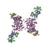
- EMDB-0246: Leucine-zippered human insulin receptor ectodomain with single bo... -
+ Open data

- Basic information
| Entry | Database: EMDB / ID: EMD-0246 | |||||||||||||||
|---|---|---|---|---|---|---|---|---|---|---|---|---|---|---|---|---|
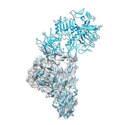
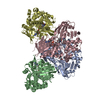
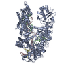
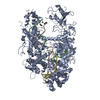
| Title | Leucine-zippered human insulin receptor ectodomain with single bound insulin - "lower" membrane-proximal part | |||||||||||||||
 Map data Map data | insulin bound to insulin receptor ectodomain - "lower" membrane-proximal part | |||||||||||||||
 Sample Sample |
| |||||||||||||||
 Keywords Keywords | insulin / insulin receptor ectodomain / signal transdution / SIGNALING PROTEIN | |||||||||||||||
| Function / homology |  Function and homology information Function and homology informationregulation of female gonad development / FCERI mediated MAPK activation / protein localization to nuclear periphery / positive regulation of meiotic cell cycle / Activation of the AP-1 family of transcription factors / negative regulation of ribosomal protein gene transcription by RNA polymerase II / positive regulation of cellular response to amino acid starvation / response to amino acid starvation / insulin-like growth factor II binding / mediator complex binding ...regulation of female gonad development / FCERI mediated MAPK activation / protein localization to nuclear periphery / positive regulation of meiotic cell cycle / Activation of the AP-1 family of transcription factors / negative regulation of ribosomal protein gene transcription by RNA polymerase II / positive regulation of cellular response to amino acid starvation / response to amino acid starvation / insulin-like growth factor II binding / mediator complex binding / positive regulation of developmental growth / insulin receptor complex / insulin-like growth factor I binding / insulin receptor activity / positive regulation of protein-containing complex disassembly / Oxidative Stress Induced Senescence / dendritic spine maintenance / adrenal gland development / insulin binding / cargo receptor activity / PTB domain binding / Signaling by Insulin receptor / IRS activation / neuronal cell body membrane / positive regulation of respiratory burst / amino acid biosynthetic process / TFIID-class transcription factor complex binding / amyloid-beta clearance / regulation of embryonic development / insulin receptor substrate binding / positive regulation of receptor internalization / positive regulation of RNA polymerase II transcription preinitiation complex assembly / positive regulation of glycogen biosynthetic process / positive regulation of transcription initiation by RNA polymerase II / Signal attenuation / protein kinase activator activity / heart morphogenesis / transport across blood-brain barrier / phosphatidylinositol 3-kinase binding / Insulin receptor recycling / insulin-like growth factor receptor binding / neuron projection maintenance / positive regulation of mitotic nuclear division / cellular response to nutrient levels / Insulin receptor signalling cascade / receptor-mediated endocytosis / dendrite membrane / positive regulation of glycolytic process / cellular response to amino acid starvation / positive regulation of D-glucose import across plasma membrane / learning / receptor protein-tyrosine kinase / male gonad development / caveola / RNA polymerase II transcription regulator complex / memory / cellular response to insulin stimulus / positive regulation of nitric oxide biosynthetic process / insulin receptor signaling pathway / late endosome / protein autophosphorylation / glucose homeostasis / amyloid-beta binding / PI5P, PP2A and IER3 Regulate PI3K/AKT Signaling / protein tyrosine kinase activity / DNA-binding transcription activator activity, RNA polymerase II-specific / transcription regulator complex / sequence-specific DNA binding / RNA polymerase II-specific DNA-binding transcription factor binding / DNA-binding transcription factor activity, RNA polymerase II-specific / positive regulation of MAPK cascade / positive regulation of canonical NF-kappaB signal transduction / positive regulation of phosphatidylinositol 3-kinase/protein kinase B signal transduction / lysosome / signaling receptor complex / endosome membrane / intracellular signal transduction / positive regulation of cell migration / RNA polymerase II cis-regulatory region sequence-specific DNA binding / DNA-binding transcription factor activity / G protein-coupled receptor signaling pathway / external side of plasma membrane / protein domain specific binding / axon / chromatin binding / positive regulation of cell population proliferation / symbiont entry into host cell / regulation of DNA-templated transcription / positive regulation of DNA-templated transcription / GTP binding / protein-containing complex binding / negative regulation of transcription by RNA polymerase II / positive regulation of transcription by RNA polymerase II / extracellular exosome / ATP binding / membrane / identical protein binding / nucleus / plasma membrane Similarity search - Function | |||||||||||||||
| Biological species |  Homo sapiens (human) / Homo sapiens (human) /  | |||||||||||||||
| Method | single particle reconstruction / cryo EM / Resolution: 4.2 Å | |||||||||||||||
 Authors Authors | Weis F / Menting JG | |||||||||||||||
| Funding support |  Australia, Australia,  United States, 4 items United States, 4 items
| |||||||||||||||
 Citation Citation | Journal: Nat Commun / Year: 2018 Title: The signalling conformation of the insulin receptor ectodomain. Authors: Felix Weis / John G Menting / Mai B Margetts / Shu Jin Chan / Yibin Xu / Norbert Tennagels / Paulus Wohlfart / Thomas Langer / Christoph W Müller / Matthias K Dreyer / Michael C Lawrence /  Abstract: Understanding the structural biology of the insulin receptor and how it signals is of key importance in the development of insulin analogs to treat diabetes. We report here a cryo-electron microscopy ...Understanding the structural biology of the insulin receptor and how it signals is of key importance in the development of insulin analogs to treat diabetes. We report here a cryo-electron microscopy structure of a single insulin bound to a physiologically relevant, high-affinity version of the receptor ectodomain, the latter generated through attachment of C-terminal leucine zipper elements to overcome the conformational flexibility associated with ectodomain truncation. The resolution of the cryo-electron microscopy maps is 3.2 Å in the insulin-binding region and 4.2 Å in the membrane-proximal region. The structure reveals how the membrane proximal domains of the receptor come together to effect signalling and how insulin's negative cooperativity of binding likely arises. Our structure further provides insight into the high affinity of certain super-mitogenic insulins. Together, these findings provide a new platform for insulin analog investigation and design. | |||||||||||||||
| History |
|
- Structure visualization
| Movie |
Movie viewer |
|---|---|
| Structure viewer | EM map: SurfViewMolmilJmol/JSmol |
| Supplemental images |
- Downloads & links
-EMDB archive
| Map data | emd_0246.map.gz | 2.5 MB | EMDB map data format | |
|---|---|---|---|---|
| Header (meta data) | emd-0246-v30.xmlemd-0246.xml | 16.7 KB 16.7 KB | Display Display | EMDB header |
| Images |  emd_0246.png emd_0246.png | 110.5 KB | ||
| Filedesc metadata | emd-0246.cif.gz | 7.2 KB | ||
| Archive directory |  http://ftp.pdbj.org/pub/emdb/structures/EMD-0246ftp://ftp.pdbj.org/pub/emdb/structures/EMD-0246 http://ftp.pdbj.org/pub/emdb/structures/EMD-0246ftp://ftp.pdbj.org/pub/emdb/structures/EMD-0246 | HTTPS FTP |
-Related structure data
| Related structure data |  6hn4MC 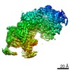 0247C  6hn5C M: atomic model generated by this map C: citing same article ( |
|---|---|
| Similar structure data |


-Links
| EMDB pages | EMDB (EBI/PDBe) / EMDataResource |
|---|---|
| Related items in Molecule of the Month |


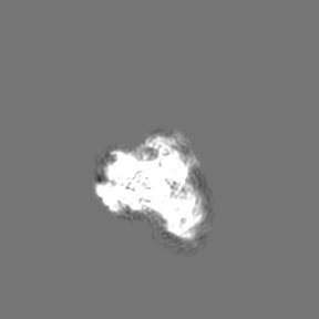
-Map
| File | Download / File: emd_0246.map.gz / Format: CCP4 / Size: 91.1 MB / Type: IMAGE STORED AS FLOATING POINT NUMBER (4 BYTES) | ||||||||||||||||||||||||||||||||||||||||||||||||||||||||||||||||||||
|---|---|---|---|---|---|---|---|---|---|---|---|---|---|---|---|---|---|---|---|---|---|---|---|---|---|---|---|---|---|---|---|---|---|---|---|---|---|---|---|---|---|---|---|---|---|---|---|---|---|---|---|---|---|---|---|---|---|---|---|---|---|---|---|---|---|---|---|---|---|
| Annotation | insulin bound to insulin receptor ectodomain - "lower" membrane-proximal part | ||||||||||||||||||||||||||||||||||||||||||||||||||||||||||||||||||||
| Projections & slices | Image control
Images are generated by Spider. | ||||||||||||||||||||||||||||||||||||||||||||||||||||||||||||||||||||
| Voxel size | X=Y=Z: 1.04 Å | ||||||||||||||||||||||||||||||||||||||||||||||||||||||||||||||||||||
| Density |
| ||||||||||||||||||||||||||||||||||||||||||||||||||||||||||||||||||||
| Symmetry | Space group: 1 | ||||||||||||||||||||||||||||||||||||||||||||||||||||||||||||||||||||
| Details | EMDB XML:
CCP4 map header:
| ||||||||||||||||||||||||||||||||||||||||||||||||||||||||||||||||||||
 Z (Sec.)
 Y (Row.)
 X (Col.)


-Supplemental data
- Sample components
-Entire : Leucine zippered human insulin receptor ectodomain (IR-A isoform,...
| Entire | Name: Leucine zippered human insulin receptor ectodomain (IR-A isoform, "deltabeta" mutant) in complex with insulin and two Fv 83-7 modules : "lower" membrane-proximal part |
|---|---|
| Components |
|
-Supramolecule #1: Leucine zippered human insulin receptor ectodomain (IR-A isoform,...
| Supramolecule | Name: Leucine zippered human insulin receptor ectodomain (IR-A isoform, "deltabeta" mutant) in complex with insulin and two Fv 83-7 modules : "lower" membrane-proximal part type: complex / ID: 1 / Parent: 0 / Macromolecule list: #1 Details: Note: Attached to the leucine-zippered insulin receptor ectodomain are two Fv 83-7 modules. One of these is present within this map volume but it is very poorly ordered and thus left ...Details: Note: Attached to the leucine-zippered insulin receptor ectodomain are two Fv 83-7 modules. One of these is present within this map volume but it is very poorly ordered and thus left completely unmodelled. See the manuscript for further details. |
|---|---|
| Source (natural) | Organism: Homo sapiens (human) |
-Macromolecule #1: Insulin receptor,Insulin receptor,General control protein GCN4
| Macromolecule | Name: Insulin receptor,Insulin receptor,General control protein GCN4 type: protein_or_peptide / ID: 1 / Number of copies: 2 / Enantiomer: LEVO / EC number: receptor protein-tyrosine kinase |
|---|---|
| Source (natural) | Organism: Strain: ATCC 204508 / S288c |
| Molecular weight | Theoretical: 106.728211 KDa |
| Recombinant expression | Organism:   Cricetulus griseus (Chinese hamster) Cricetulus griseus (Chinese hamster) |
| Sequence | String: HLYPGEVCPG MDIRNNLTRL HELENCSVIE GHLQILLMFK TRPEDFRDLS FPKLIMITDY LLLFRVYGLE SLKDLFPNLT VIRGSRLFF NYALVIFEMV HLKELGLYNL MNITRGSVRI EKNNELCYLA TIDWSRILDS VEDNHIVLNK DDNEECGDIC P GTAKGKTN ...String: HLYPGEVCPG MDIRNNLTRL HELENCSVIE GHLQILLMFK TRPEDFRDLS FPKLIMITDY LLLFRVYGLE SLKDLFPNLT VIRGSRLFF NYALVIFEMV HLKELGLYNL MNITRGSVRI EKNNELCYLA TIDWSRILDS VEDNHIVLNK DDNEECGDIC P GTAKGKTN CPATVINGQF VERCWTHSHC QKVCPTICKS HGCTAEGLCC HSECLGNCSQ PDDPTKCVAC RNFYLDGRCV ET CPPPYYH FQDWRCVNFS FCQDLHHKCK NSRRQGCHQY VIHNNKCIPE CPSGYTMNSS NLLCTPCLGP CPKVCHLLEG EKT IDSVTS AQELRGCTVI NGSLIINIRG GNNLAAELEA NLGLIEEISG YLKIRRSYAL VSLSFFRKLR LIRGETLEIG NYSF YALDN QNLRQLWDWS KHNLTTTQGK LFFHYNPKLC LSEIHKMEEV SGTKGRQERN DIALKTNGDK ASCENELLKF SYIRT SFDK ILLRWEPYWP PDFRDLLGFM LFYKEAPYQN VTEFDGQDAC GSNSWTVVDI DPPLRSNDPK SQNHPGWLMR GLKPWT QYA IFVKTLVTFS DERRTYGAKS DIIYVQTDAT NPSVPLDPIS VSNSSSQIIL KWKPPSDPNG NITHYLVFWE RQAEDSE LF ELDYCLKGLK LPSRTWSPPF ESEDSQKHNQ SEYEDSAGEC CSCPKTDSQI LKELEESSFR KTFEDYLHNV VFVPRPSR K RRSLGDVGNA GNNEEHRPFE KVVNKESLVI SGLRHFTGYR IELQACNQDT PEERCSVAAY VSARTMPEAK ADDIVGPVT HEIFENNVVH LMWQEPKEPN GLIVLYEVSY RRYGDEELHL CVSRKHFALE RGCRLRGLSP GNYSVRIRAT SLAGNGSWTE PTYFYVTDY LDVPSNIARM KQLEDKVEEL LSKNYHLENE VARLKKLVGE R UniProtKB: Insulin receptor, Insulin receptor, General control transcription factor GCN4 |
-Macromolecule #2: 2-acetamido-2-deoxy-beta-D-glucopyranose
| Macromolecule | Name: 2-acetamido-2-deoxy-beta-D-glucopyranose / type: ligand / ID: 2 / Number of copies: 6 / Formula: NAG |
|---|---|
| Molecular weight | Theoretical: 221.208 Da |
| Chemical component information |  ChemComp-NAG: |
-Experimental details
-Structure determination
| Method | cryo EM |
|---|---|
 Processing Processing | single particle reconstruction |
| Aggregation state | particle |
-Sample preparation
| Concentration | 0.094 mg/mL | |||||||||
|---|---|---|---|---|---|---|---|---|---|---|
| Buffer | pH: 7.5 Component:
| |||||||||
| Vitrification | Cryogen name: ETHANE / Chamber humidity: 100 % / Chamber temperature: 283.15 K / Instrument: FEI VITROBOT MARK IV |
- Electron microscopy
| Microscope | FEI TITAN KRIOS |
|---|---|
| Specialist optics | Energy filter - Name: GIF Quantum LS / Energy filter - Slit width: 20 eV |
| Image recording | Film or detector model: GATAN K2 SUMMIT (4k x 4k) / Detector mode: SUPER-RESOLUTION / Digitization - Frames/image: 1-20 / Number grids imaged: 1 / Number real images: 2287 / Average exposure time: 16.0 sec. / Average electron dose: 1.85 e/Å2 |
| Electron beam | Acceleration voltage: 300 kV / Electron source:  FIELD EMISSION GUN FIELD EMISSION GUN |
| Electron optics | C2 aperture diameter: 50.0 µm / Illumination mode: FLOOD BEAM / Imaging mode: BRIGHT FIELD / Cs: 2.7 mm / Nominal defocus max: 2.5 µm / Nominal defocus min: 1.0 µm / Nominal magnification: 130000 |
| Sample stage | Specimen holder model: FEI TITAN KRIOS AUTOGRID HOLDER / Cooling holder cryogen: NITROGEN |
| Experimental equipment |  Model: Titan Krios / Image courtesy: FEI Company |
+Image processing
-Atomic model buiding 1
| Initial model | PDB ID: Chain - Source name: PDB / Chain - Initial model type: experimental model |
|---|---|
| Refinement | Space: REAL / Protocol: OTHER / Target criteria: Cross-correlation coefficient |
| Output model | PDB-6hn4: |
