 Movie
Movie Controller

[English] 日本語
 Yorodumi
- EMDB-0098: tRNA translocation by the eukaryotic 80S ribosome and the impact ... -
+ Open data

- Basic information
| Entry | Database: EMDB / ID: EMD-0098 | ||||||||||||
|---|---|---|---|---|---|---|---|---|---|---|---|---|---|
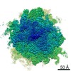
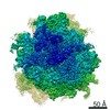
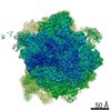
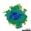
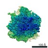
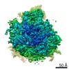
| Title | tRNA translocation by the eukaryotic 80S ribosome and the impact of GTP hydrolysis, Translocation-intermediate-POST-1 (TI-POST-1) | ||||||||||||
 Map data Map data | |||||||||||||
 Sample Sample |
| ||||||||||||
| Function / homology |  Function and homology information Function and homology information Ribosomal protein S12e signature. / 40S Ribosomal protein S10 / Plectin/S10, N-terminal / Plectin/S10 domain / Ribosomal protein L1 signature. / Ribosomal protein S17e signature. / Ribosomal protein L44e signature. / Ribosomal protein L27e signature. / Ribosomal protein L10e signature. / Ribosomal protein S7e signature. ...Ribosomal protein S12e signature. / 40S Ribosomal protein S10 / Plectin/S10, N-terminal / Plectin/S10 domain / Ribosomal protein L1 signature. / Ribosomal protein S17e signature. / Ribosomal protein L44e signature. / Ribosomal protein L27e signature. / Ribosomal protein L10e signature. / Ribosomal protein S7e signature. / Ribosomal protein S3Ae signature. / Ribosomal protein L24e signature. / Ribosomal protein L39e signature. / Ribosomal protein S6e signature. / Ribosomal protein S28e signature. / Ribosomal protein L31e signature. / Ribosomal protein L32e signature. / Ribosomal protein L15e signature. / Ribosomal protein L21e signature. / Ribosomal protein L37e signature. / Ribosomal protein L11 signature. / Ribosomal protein S14 signature. / Ribosomal protein L5 signature. / Ribosomal protein S7 signature. / Ribosomal protein S17 signature. / Ribosomal protein S13 signature. / Ribosomal protein S13 family profile. / Ribosomal protein S8 signature. / Ribosomal protein S4 signature. / Ribosomal protein S15 signature. / S4 RNA-binding domain profile. / Ribosomal protein L14 signature. / Ubiquitin domain profile. / Ribosomal protein S12 signature. / Ribosomal protein L24 signature. / Trp-Asp (WD) repeats signature. / Trp-Asp (WD) repeats profile. / Trp-Asp (WD) repeats circular profile. / Winged helix-like DNA-binding domain superfamily Ribosomal protein S12e signature. / 40S Ribosomal protein S10 / Plectin/S10, N-terminal / Plectin/S10 domain / Ribosomal protein L1 signature. / Ribosomal protein S17e signature. / Ribosomal protein L44e signature. / Ribosomal protein L27e signature. / Ribosomal protein L10e signature. / Ribosomal protein S7e signature. ...Ribosomal protein S12e signature. / 40S Ribosomal protein S10 / Plectin/S10, N-terminal / Plectin/S10 domain / Ribosomal protein L1 signature. / Ribosomal protein S17e signature. / Ribosomal protein L44e signature. / Ribosomal protein L27e signature. / Ribosomal protein L10e signature. / Ribosomal protein S7e signature. / Ribosomal protein S3Ae signature. / Ribosomal protein L24e signature. / Ribosomal protein L39e signature. / Ribosomal protein S6e signature. / Ribosomal protein S28e signature. / Ribosomal protein L31e signature. / Ribosomal protein L32e signature. / Ribosomal protein L15e signature. / Ribosomal protein L21e signature. / Ribosomal protein L37e signature. / Ribosomal protein L11 signature. / Ribosomal protein S14 signature. / Ribosomal protein L5 signature. / Ribosomal protein S7 signature. / Ribosomal protein S17 signature. / Ribosomal protein S13 signature. / Ribosomal protein S13 family profile. / Ribosomal protein S8 signature. / Ribosomal protein S4 signature. / Ribosomal protein S15 signature. / S4 RNA-binding domain profile. / Ribosomal protein L14 signature. / Ubiquitin domain profile. / Ribosomal protein S12 signature. / Ribosomal protein L24 signature. / Trp-Asp (WD) repeats signature. / Trp-Asp (WD) repeats profile. / Trp-Asp (WD) repeats circular profile. / Winged helix-like DNA-binding domain superfamilySimilarity search - Domain/homology | ||||||||||||
| Biological species |  Oryctolagus cuniculus (rabbit) / Oryctolagus cuniculus (rabbit) /  Enterobacteria phage SP6 (virus) / Enterobacteria phage SP6 (virus) /  Saccharomyces cerevisiae (brewer's yeast) / Rabbit (rabbit) Saccharomyces cerevisiae (brewer's yeast) / Rabbit (rabbit) | ||||||||||||
| Method | single particle reconstruction / cryo EM / Resolution: 3.6 Å | ||||||||||||
 Authors Authors | Flis J / Holm M / Rundlet EJ / Loerke J / Hilal T / Dabrowski M / Buerger J / Mielke T / Blanchard SC / Spahn CMT / Budkevich TV | ||||||||||||
| Funding support |  Germany, Germany,  United States, 3 items United States, 3 items
| ||||||||||||
 Citation Citation | Journal: Cell Rep / Year: 2018 Title: tRNA Translocation by the Eukaryotic 80S Ribosome and the Impact of GTP Hydrolysis. Authors: Julia Flis / Mikael Holm / Emily J Rundlet / Justus Loerke / Tarek Hilal / Marylena Dabrowski / Jörg Bürger / Thorsten Mielke / Scott C Blanchard / Christian M T Spahn / Tatyana V Budkevich / Abstract: Translocation moves the tRNA⋅mRNA module directionally through the ribosome during the elongation phase of protein synthesis. Although translocation is known to entail large conformational changes ...Translocation moves the tRNA⋅mRNA module directionally through the ribosome during the elongation phase of protein synthesis. Although translocation is known to entail large conformational changes within both the ribosome and tRNA substrates, the orchestrated events that ensure the speed and fidelity of this critical aspect of the protein synthesis mechanism have not been fully elucidated. Here, we present three high-resolution structures of intermediates of translocation on the mammalian ribosome where, in contrast to bacteria, ribosomal complexes containing the translocase eEF2 and the complete tRNA⋅mRNA module are trapped by the non-hydrolyzable GTP analog GMPPNP. Consistent with the observed structures, single-molecule imaging revealed that GTP hydrolysis principally facilitates rate-limiting, final steps of translocation, which are required for factor dissociation and which are differentially regulated in bacterial and mammalian systems by the rates of deacyl-tRNA dissociation from the E site. | ||||||||||||
| History |
|
- Structure visualization
| Movie |
Movie viewer |
|---|---|
| Structure viewer | EM map: SurfViewMolmilJmol/JSmol |
| Supplemental images |
- Downloads & links
-EMDB archive
| Map data | emd_0098.map.gz | 230.3 MB | EMDB map data format | |
|---|---|---|---|---|
| Header (meta data) | emd-0098-v30.xmlemd-0098.xml | 105.4 KB 105.4 KB | Display Display | EMDB header |
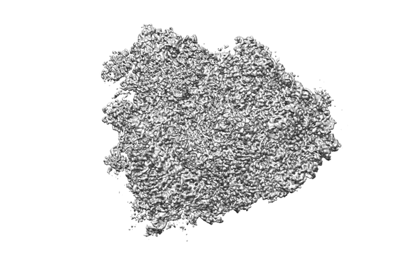
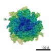
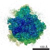
| Images |  emd_0098.png emd_0098.png | 72.2 KB | ||
| Archive directory |  http://ftp.pdbj.org/pub/emdb/structures/EMD-0098ftp://ftp.pdbj.org/pub/emdb/structures/EMD-0098 http://ftp.pdbj.org/pub/emdb/structures/EMD-0098ftp://ftp.pdbj.org/pub/emdb/structures/EMD-0098 | HTTPS FTP |
-Related structure data
| Related structure data |  6gz3MC 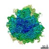 0099C 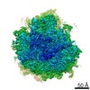 0100C 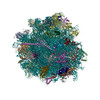 6gz4C 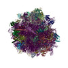 6gz5C M: atomic model generated by this map C: citing same article ( |
|---|---|
| Similar structure data |

-Links
| EMDB pages | EMDB (EBI/PDBe) / EMDataResource |
|---|---|
| Related items in Molecule of the Month |


-Map
| File | Download / File: emd_0098.map.gz / Format: CCP4 / Size: 253.4 MB / Type: IMAGE STORED AS FLOATING POINT NUMBER (4 BYTES) | ||||||||||||||||||||||||||||||||||||||||||||||||||||||||||||||||||||
|---|---|---|---|---|---|---|---|---|---|---|---|---|---|---|---|---|---|---|---|---|---|---|---|---|---|---|---|---|---|---|---|---|---|---|---|---|---|---|---|---|---|---|---|---|---|---|---|---|---|---|---|---|---|---|---|---|---|---|---|---|---|---|---|---|---|---|---|---|---|
| Voxel size | X=Y=Z: 0.975 Å | ||||||||||||||||||||||||||||||||||||||||||||||||||||||||||||||||||||
| Density |
| ||||||||||||||||||||||||||||||||||||||||||||||||||||||||||||||||||||
| Symmetry | Space group: 1 | ||||||||||||||||||||||||||||||||||||||||||||||||||||||||||||||||||||
| Details | EMDB XML:
CCP4 map header:
| ||||||||||||||||||||||||||||||||||||||||||||||||||||||||||||||||||||
-Supplemental data
- Sample components
+Entire : translocation intermediate TI-POST-1
+Supramolecule #1: translocation intermediate TI-POST-1
+Supramolecule #2: Ribosome
+Supramolecule #3: mRNA
+Supramolecule #4: tRNA
+Macromolecule #1: 28S ribosomal RNA
+Macromolecule #2: ap/P-site tRNA
+Macromolecule #3: mRNA
+Macromolecule #4: pe/E-site-tRNA
+Macromolecule #5: 18S ribosomal RNA
+Macromolecule #39: 5.8S ribosomal RNA
+Macromolecule #40: 5S ribosomal RNA
+Macromolecule #6: ribosomal protein uS3
+Macromolecule #7: Ribosomal protein S5
+Macromolecule #8: ribosomal protein eS10
+Macromolecule #9: 40S ribosomal protein S12
+Macromolecule #10: ribosomal protein uS19
+Macromolecule #11: ribosomal protein uS9
+Macromolecule #12: ribosomal protein eS17
+Macromolecule #13: ribosomal protein uS13
+Macromolecule #14: ribosomal protein eS19
+Macromolecule #15: ribosomal protein uS10
+Macromolecule #16: ribosomal protein eS25
+Macromolecule #17: Ribosomal protein S28
+Macromolecule #18: ribosomal protein uS14
+Macromolecule #19: Ribosomal protein S27a
+Macromolecule #20: ribosomal protein RACK 1
+Macromolecule #21: ribosomal protein uS2
+Macromolecule #22: 40S ribosomal protein S3a
+Macromolecule #23: ribosomal protein uS5
+Macromolecule #24: ribosomal protein eS4
+Macromolecule #25: 40S ribosomal protein S6
+Macromolecule #26: 40S ribosomal protein S7
+Macromolecule #27: ribosomal protein eS8
+Macromolecule #28: Ribosomal protein S9 (Predicted)
+Macromolecule #29: Ribosomal protein S11
+Macromolecule #30: ribosomal protein uS15
+Macromolecule #31: ribosomal protein uS11
+Macromolecule #32: ribosomal protein eS21
+Macromolecule #33: Ribosomal protein S15a
+Macromolecule #34: Ribosomal protein S23
+Macromolecule #35: ribosomal protein eS24
+Macromolecule #36: ribosomal protein eS26
+Macromolecule #37: ribosomal protein eS27
+Macromolecule #38: ribosomal protein eS30
+Macromolecule #41: Ribosomal protein L8
+Macromolecule #42: ribosomal protein uL3
+Macromolecule #43: ribosomal protein uL4
+Macromolecule #44: ribosomal protein uL18
+Macromolecule #45: ribosomal protein eL6
+Macromolecule #46: ribosomal protein uL30
+Macromolecule #47: ribosomal protein eL8
+Macromolecule #48: ribosomal protein uL6
+Macromolecule #49: Ribosomal protein L10 (Predicted)
+Macromolecule #50: Ribosomal protein L11
+Macromolecule #51: ribosomal protein eL13
+Macromolecule #52: ribosomal protein eL14
+Macromolecule #53: Ribosomal protein L15
+Macromolecule #54: ribosomal protein uL13
+Macromolecule #55: ribosomal protein uL22
+Macromolecule #56: ribosomal protein eL18
+Macromolecule #57: ribosomal protein eL19
+Macromolecule #58: ribosomal protein eL20
+Macromolecule #59: ribosomal protein eL21
+Macromolecule #60: ribosomal protein eL22
+Macromolecule #61: Ribosomal protein L23
+Macromolecule #62: ribosomal protein eL24
+Macromolecule #63: ribosomal protein uL23
+Macromolecule #64: Ribosomal protein L26
+Macromolecule #65: 60S ribosomal protein L27
+Macromolecule #66: ribosomal protein uL15
+Macromolecule #67: ribosomal protein eL29
+Macromolecule #68: ribosomal protein eL30
+Macromolecule #69: ribosomal protein eL31
+Macromolecule #70: ribosomal protein eL32
+Macromolecule #71: ribosomal protein eL33
+Macromolecule #72: ribosomal protein eL34
+Macromolecule #73: ribosomal protein uL29
+Macromolecule #74: ribosomal protein eL36
+Macromolecule #75: Ribosomal protein L37
+Macromolecule #76: ribosomal protein eL38
+Macromolecule #77: ribosomal protein eL39
+Macromolecule #78: ribosomal protein eL40
+Macromolecule #79: ribosomal protein eL41
+Macromolecule #80: ribosomal protein eL42
+Macromolecule #81: ribosomal protein eL43
+Macromolecule #82: ribosomal protein eL28
+Macromolecule #83: Ribosomal protein
+Macromolecule #84: Ribosomal protein L12
+Macromolecule #85: ribosomal protein uL10
+Macromolecule #86: eukaryotic elongation factor 2 (eEF2)
+Macromolecule #87: MAGNESIUM ION
+Macromolecule #88: ZINC ION
+Macromolecule #89: PHOSPHOAMINOPHOSPHONIC ACID-GUANYLATE ESTER

-Experimental details
-Structure determination
| Method | cryo EM |
|---|---|
 Processing Processing | single particle reconstruction |
| Aggregation state | particle |
-Sample preparation
| Buffer | pH: 7.5 |
|---|---|
| Vitrification | Cryogen name: ETHANE |
- Electron microscopy
| Microscope | FEI POLARA 300 |
|---|---|
| Electron beam | Acceleration voltage: 300 kV / Electron source: FIELD EMISSION GUN |
| Electron optics | Illumination mode: FLOOD BEAM / Imaging mode: BRIGHT FIELDBright-field microscopy |
| Image recording | Film or detector model: GATAN K2 SUMMIT (4k x 4k) / Detector mode: SUPER-RESOLUTION / Average exposure time: 5.0 sec. / Average electron dose: 30.0 e/Å2 |
| Experimental equipment |  Model: Tecnai Polara / Image courtesy: FEI Company |
-Image processing
| Particle selection | Number selected: 270000 |
|---|---|
| Startup model | Type of model: OTHER / Details: filtered empty 80S |
| Initial angle assignment | Type: PROJECTION MATCHING |
| Final angle assignment | Type: PROJECTION MATCHING |
| Final reconstruction | Applied symmetry - Point group: C1 (asymmetric) / Resolution.type: BY AUTHOR / Resolution: 3.6 Å / Resolution method: FSC 0.143 CUT-OFF / Number images used: 32386 |
-Atomic model buiding 1
| Refinement | Space: REAL / Protocol: RIGID BODY FIT |
|---|---|
| Output model | PDB-6gz3: |
