 Movie
Movie Controller
 Structure viewers
 About Yorodumi Papers


+Search query
-Structure paper
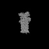
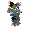
| Title | Structural insights into the ubiquitin-independent midnolin-proteasome pathway. |
|---|---|
| Journal, issue, pages | Proc Natl Acad Sci U S A, Vol. 122, Issue 19, Page e2505345122, Year 2025 |
| Publish date | May 13, 2025 |
 Authors Authors | Nagesh Peddada / Xue Zhong / Yan Yin / Danielle Renee Lazaro / Jianhui Wang / Stephen Lyon / Jin Huk Choi / Xiao-Chen Bai / Eva Marie Y Moresco / Bruce Beutler /  |
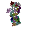
| PubMed Abstract | The protein midnolin (MIDN) augments proteasome activity in lymphocytes and dramatically facilitates the survival and proliferation of B-lymphoid malignancies. MIDN binds both to proteasomes and to ...The protein midnolin (MIDN) augments proteasome activity in lymphocytes and dramatically facilitates the survival and proliferation of B-lymphoid malignancies. MIDN binds both to proteasomes and to substrates, but the mode of interaction with the proteasome is unknown, and the mechanism by which MIDN facilitates substrate degradation in a ubiquitin-independent manner is incompletely understood. Here, we present cryoelectron microscopy (cryo-EM) structures of the substrate-engaged, MIDN-bound human proteasome in two conformational states. MIDN induces proteasome conformations similarly to ubiquitinated substrates by using its ubiquitin-like domain to bind to the deubiquitinase RPN11 (PSMD14). By simultaneously binding to RPN1 (PSMD2) with its C-terminal α-helix, MIDN positions its substrate-carrying Catch domain above the proteasome ATPase channel through which substrates are translocated before degradation. Our findings suggest that both ubiquitin-like domain and C-terminal α-helix must bind to the proteasome for MIDN to stimulate proteasome activity. |
 External links External links | Proc Natl Acad Sci U S A / PubMed:40339123 / PubMed Central |
| Methods | EM (single particle) |
| Resolution | 2.8 - 3.84 Å |
| Structure data | 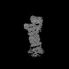 EMDB-49497: Consensus map of MIDN-bound 26S proteasome, EB-state 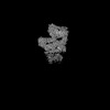 EMDB-49498: Locally Refined map of RP(19S) in substrate-engaged MIDN-bound 26S Proteasome, EB-MIDN state  EMDB-49499: Locally refined map of RPN1-MIDN_alphaHelix-C  EMDB-49500: Locally refined map of RPN11-MIDN_UBL domain 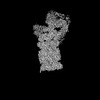 EMDB-49501: Consensus map of 26S proteasome bound to MIDN, EB-MIDN_UBL state 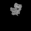 EMDB-49502: Locally refined map of RP(19S) of MIDN-bound 26S proteasome in EB-MIDN_UBL state 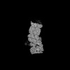 EMDB-49503: Consensus map of substrate-free 26S proteasome in presence MG-132 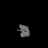 EMDB-49504: Focused map of RP (19S) substrate-free MIDN-free 26S proteasome, SA-like state with MG-132  EMDB-49505: Consensus map of substrate engaged MIDN-bound 26S proteasome, ED-state  EMDB-49506: Locally Refined map of RP(19S) in substrate-engaged MIDN-bound 26S Proteasome, ED-MIDN state EMDB-49507, PDB-9nkf: EMDB-49508, PDB-9nkg: EMDB-49509, PDB-9nki: EMDB-49510, PDB-9nkj: |
| Chemicals |  ChemComp-ATP:  ChemComp-MG:  ChemComp-ADP:  ChemComp-LDZ:  ChemComp-ZN: |
| Source |
|
 Keywords Keywords | IMMUNE SYSTEM / Apo state / proteasome / Midnolin / substrate-free 26S / MG-132 / Ubiquitin-indepedent / MIDN-bound 26S complex / MIDN-UBL / 26S complex / ED-state |
 homo sapiens (human)