Movie
Movie Controller
 Structure viewers
 About Yorodumi Papers


+Search query
-Structure paper
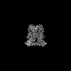
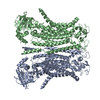
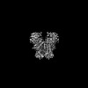
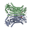
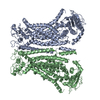
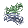
| Title | Structural basis for the activation of the lipid scramblase TMEM16F. |
|---|---|
| Journal, issue, pages | Nat Commun, Vol. 13, Issue 1, Page 6692, Year 2022 |
| Publish date | Nov 5, 2022 |
 Authors Authors | Melanie Arndt / Carolina Alvadia / Monique S Straub / Vanessa Clerico Mosina / Cristina Paulino / Raimund Dutzler /   |
| PubMed Abstract | TMEM16F, a member of the conserved TMEM16 family, plays a central role in the initiation of blood coagulation and the fusion of trophoblasts. The protein mediates passive ion and lipid transport in ...TMEM16F, a member of the conserved TMEM16 family, plays a central role in the initiation of blood coagulation and the fusion of trophoblasts. The protein mediates passive ion and lipid transport in response to an increase in intracellular Ca. However, the mechanism of how the protein facilitates both processes has remained elusive. Here we investigate the basis for TMEM16F activation. In a screen of residues lining the proposed site of conduction, we identify mutants with strongly activating phenotype. Structures of these mutants determined herein by cryo-electron microscopy show major rearrangements leading to the exposure of hydrophilic patches to the membrane, whose distortion facilitates lipid diffusion. The concomitant opening of a pore promotes ion conduction in the same protein conformation. Our work has revealed a mechanism that is distinct for this branch of the family and that will aid the development of a specific pharmacology for a promising drug target. |
 External links External links | Nat Commun / PubMed:36335104 / PubMed Central |
| Methods | EM (single particle) |
| Resolution | 2.93 - 3.49 Å |
| Structure data | EMDB-15913, PDB-8b8g: EMDB-15914, PDB-8b8j: EMDB-15916, PDB-8b8k: EMDB-15917, PDB-8b8m: EMDB-15919, PDB-8b8q: EMDB-15958, PDB-8bc0: EMDB-15959, PDB-8bc1: |
| Chemicals |  ChemComp-CA:  ChemComp-P1O: |
| Source |
|
 Keywords Keywords | MEMBRANE PROTEIN / Lipid Transport / Lipid Scrambling / Ion Channel / Plasma Membrane / Blood Clotting / Exocytosis / Membrane Fusion / Lipid Scramblase / Blood Coagulation / Viral Entry / Cell Fusion |