Movie
Movie Controller


+ Open data

- Basic information
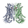
| Entry | Database: PDB / ID: 8b8q | ||||||
|---|---|---|---|---|---|---|---|
| Title | Structure of mTMEM16F in lipid Nanodiscs in the presence of Ca2+ | ||||||
 Components Components | Anoctamin-6 | ||||||
 Keywords Keywords | MEMBRANE PROTEIN / Lipid Transport / Lipid Scramblase / Ion Channel / Blood Coagulation / Membrane Fusion | ||||||
| Function / homology |  Function and homology information Function and homology informationcalcium activated galactosylceramide scrambling / phosphatidylserine exposure on blood platelet / calcium activated phosphatidylserine scrambling / calcium activated phosphatidylcholine scrambling / positive regulation of potassium ion export across plasma membrane / positive regulation of monoatomic ion transmembrane transport / calcium activated phospholipid scrambling / activation of blood coagulation via clotting cascade / purinergic nucleotide receptor signaling pathway / bone mineralization involved in bone maturation ...calcium activated galactosylceramide scrambling / phosphatidylserine exposure on blood platelet / calcium activated phosphatidylserine scrambling / calcium activated phosphatidylcholine scrambling / positive regulation of potassium ion export across plasma membrane / positive regulation of monoatomic ion transmembrane transport / calcium activated phospholipid scrambling / activation of blood coagulation via clotting cascade / purinergic nucleotide receptor signaling pathway / bone mineralization involved in bone maturation / phospholipid scramblase activity / cholinergic synapse / intracellularly calcium-gated chloride channel activity / pore complex assembly / negative regulation of cell volume / plasma membrane phospholipid scrambling / bleb assembly / voltage-gated monoatomic ion channel activity / positive regulation of phagocytosis, engulfment / Stimuli-sensing channels / voltage-gated chloride channel activity / positive regulation of monocyte chemotaxis / calcium-activated cation channel activity / chloride transport / phospholipid translocation / dendritic cell chemotaxis / chloride channel activity / regulation of postsynaptic membrane potential / positive regulation of endothelial cell apoptotic process / positive regulation of bone mineralization / chloride channel complex / Neutrophil degranulation / chloride transmembrane transport / sodium ion transmembrane transport / synaptic membrane / calcium ion transmembrane transport / blood coagulation / positive regulation of apoptotic process / protein homodimerization activity / metal ion binding / identical protein binding / plasma membrane / cytosol Similarity search - Function | ||||||
| Biological species |  | ||||||
| Method | ELECTRON MICROSCOPY / single particle reconstruction / cryo EM / Resolution: 2.94 Å | ||||||
 Authors Authors | Arndt, M. / Alvadia, C. / Straub, M.S. / Clerico-Mosina, V. / Paulino, C. / Dutzler, R. | ||||||
| Funding support | European Union, 1items
| ||||||
 Citation Citation | Journal: Nat Commun / Year: 2022 Title: Structural basis for the activation of the lipid scramblase TMEM16F. Authors: Melanie Arndt / Carolina Alvadia / Monique S Straub / Vanessa Clerico Mosina / Cristina Paulino / Raimund Dutzler /   Abstract: TMEM16F, a member of the conserved TMEM16 family, plays a central role in the initiation of blood coagulation and the fusion of trophoblasts. The protein mediates passive ion and lipid transport in ...TMEM16F, a member of the conserved TMEM16 family, plays a central role in the initiation of blood coagulation and the fusion of trophoblasts. The protein mediates passive ion and lipid transport in response to an increase in intracellular Ca. However, the mechanism of how the protein facilitates both processes has remained elusive. Here we investigate the basis for TMEM16F activation. In a screen of residues lining the proposed site of conduction, we identify mutants with strongly activating phenotype. Structures of these mutants determined herein by cryo-electron microscopy show major rearrangements leading to the exposure of hydrophilic patches to the membrane, whose distortion facilitates lipid diffusion. The concomitant opening of a pore promotes ion conduction in the same protein conformation. Our work has revealed a mechanism that is distinct for this branch of the family and that will aid the development of a specific pharmacology for a promising drug target. | ||||||
| History |
|
- Structure visualization
| Structure viewer | Molecule: MolmilJmol/JSmol |
|---|
- Downloads & links
-Download
| PDBx/mmCIF format | 8b8q.cif.gz | 297.7 KB | Display | PDBx/mmCIF format |
|---|---|---|---|---|
| PDB format | pdb8b8q.ent.gz | 233.9 KB | Display | PDB format |
| PDBx/mmJSON format | 8b8q.json.gz | Tree view | PDBx/mmJSON format | |
| Others |  Other downloads Other downloads |
-Validation report
| Arichive directory | https://data.pdbj.org/pub/pdb/validation_reports/b8/8b8qftp://data.pdbj.org/pub/pdb/validation_reports/b8/8b8q | HTTPS FTP |
|---|
-Related structure data
| Related structure data |  15919MC 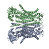 8b8gC  8b8jC 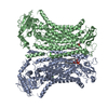 8b8kC 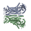 8b8mC 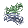 8bc0C  8bc1C M: map data used to model this data C: citing same article ( |
|---|---|
| Similar structure data |


-Links
 PDBj
PDBj


- Assembly
| Deposited unit | 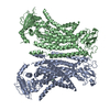
|
|---|---|
| 1 |
|
-Components
| #1: Protein | Mass: 113454.602 Da / Num. of mol.: 2 / Mutation: F518H Source method: isolated from a genetically manipulated source Source: (gene. exp.)  Homo sapiens (human) / References: UniProt: Q6P9J9 Homo sapiens (human) / References: UniProt: Q6P9J9#2: Chemical | ChemComp-CA /   Mass: 40.078 Da / Num. of mol.: 6 / Source method: obtained synthetically / Formula: Ca Mass: 40.078 Da / Num. of mol.: 6 / Source method: obtained synthetically / Formula: CaHas protein modification | Y | |
|---|
-Experimental details
-Experiment
| Experiment | Method: ELECTRON MICROSCOPY |
|---|---|
| EM experiment | Aggregation state: PARTICLE / 3D reconstruction method: single particle reconstruction |
- Sample preparation
| Component | Name: mTMEM16F Homodimer bound to Ca2+ / Type: COMPLEX / Entity ID: #1 / Source: RECOMBINANT |
|---|---|
| Molecular weight | Experimental value: NO |
| Source (natural) | Organism: |
| Source (recombinant) | Organism: Homo sapiens (human) |
| Buffer solution | pH: 7.5 |
| Specimen | Embedding applied: NO / Shadowing applied: NO / Staining applied: NO / Vitrification applied: YES |
| Vitrification | Cryogen name: ETHANE-PROPANE |
- Electron microscopy imaging
| Experimental equipment |  Model: Titan Krios / Image courtesy: FEI Company |
|---|---|
| Microscopy | Model: FEI TITAN KRIOS |
| Electron gun | Electron source:  FIELD EMISSION GUN / Accelerating voltage: 300 kV / Illumination mode: OTHER FIELD EMISSION GUN / Accelerating voltage: 300 kV / Illumination mode: OTHER |
| Electron lens | Mode: BRIGHT FIELD / Nominal defocus max: 2400 nm / Nominal defocus min: 1000 nm / Cs: 2.7 mm |
| Image recording | Electron dose: 66.6 e/Å2 / Film or detector model: GATAN K3 (6k x 4k) Details: 3 Datasets were collected from differenct grids. Eposure time was between 66.6 e-/A2 for GO grids, 84.2 e-/A2 for UltrAu foil grids, and 76 e-/A2 for UltrAu foil grids with 20deg tilt collection |
- Processing
| Software | Name: PHENIX / Version: 1.20.1_4487: / Classification: refinement | ||||||||||||||||||||||||
|---|---|---|---|---|---|---|---|---|---|---|---|---|---|---|---|---|---|---|---|---|---|---|---|---|---|
| EM software |
| ||||||||||||||||||||||||
| CTF correction | Type: PHASE FLIPPING AND AMPLITUDE CORRECTION | ||||||||||||||||||||||||
| Symmetry | Point symmetry: C1 (asymmetric) | ||||||||||||||||||||||||
| 3D reconstruction | Resolution: 2.94 Å / Resolution method: FSC 0.143 CUT-OFF / Num. of particles: 282719 / Symmetry type: POINT | ||||||||||||||||||||||||
| Atomic model building | B value: 85 / Protocol: FLEXIBLE FIT / Space: REAL | ||||||||||||||||||||||||
| Atomic model building | PDB-ID: 6QP6 Accession code: 6QP6 / Source name: PDB / Type: experimental model | ||||||||||||||||||||||||
| Refine LS restraints |
|