PDB-6dt3: 1.2 Angstrom Resolution Crystal Structure of Nucleoside Triphosphatase NudI from Klebsiella pneumoniae in Complex with HEPES Method: X-RAY DIFFRACTION / Resolution: 1.2 Å
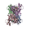
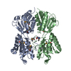
PDB-6dux: 2.25 Angstrom Resolution Crystal Structure of 6-phospho-alpha-glucosidase from Klebsiella pneumoniae in Complex with NAD. Method: X-RAY DIFFRACTION / Resolution: 2.25 Å
PDB-6dvv: 2.25 Angstrom Resolution Crystal Structure of 6-phospho-alpha-glucosidase from Klebsiella pneumoniae in Complex with NAD and Mn2+. Method: X-RAY DIFFRACTION / Resolution: 2.25 Å
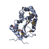
PDB-6dxn: 1.95 Angstrom Resolution Crystal Structure of DsbA Disulfide Interchange Protein from Klebsiella pneumoniae. Method: X-RAY DIFFRACTION / Resolution: 1.95 Å
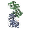
PDB-6e85: 1.25 Angstrom Resolution Crystal Structure of 4-hydroxythreonine-4-phosphate Dehydrogenase from Klebsiella pneumoniae. Method: X-RAY DIFFRACTION / Resolution: 1.25 Å
PDB-6nau: 1.55 Angstrom Resolution Crystal Structure of 6-phosphogluconolactonase from Klebsiella pneumoniae Method: X-RAY DIFFRACTION / Resolution: 1.55 Å
PDB-6nbg: 2.05 Angstrom Resolution Crystal Structure of Hypothetical Protein KP1_5497 from Klebsiella pneumoniae. Method: X-RAY DIFFRACTION / Resolution: 2.05 Å
PDB-6ndi: Crystal Structure of the Sugar Binding Domain of LacI Family Protein from Klebsiella pneumoniae Method: X-RAY DIFFRACTION / Resolution: 2.6 Å
PDB-6wn5: 1.52 Angstrom Resolution Crystal Structure of Transcriptional Regulator HdfR from Klebsiella pneumoniae Method: X-RAY DIFFRACTION / Resolution: 1.52 Å
PDB-6wn8: 2.70 Angstrom Resolution Crystal Structure of Uracil Phosphoribosyl Transferase from Klebsiella pneumoniae Method: X-RAY DIFFRACTION / Resolution: 2.7 Å
PDB-6x1l: The crystal structure of a functional uncharacterized protein KP1_0663 from Klebsiella pneumoniae subsp. pneumoniae NTUH-K2044 Method: X-RAY DIFFRACTION / Resolution: 2 Å
PDB-7rjj: Crystal Structure of the Peptidoglycan Binding Domain of the Outer Membrane Protein (OmpA) from Klebsiella pneumoniae with bound D-alanine Method: X-RAY DIFFRACTION / Resolution: 1.88 Å
PDB-7tl5: Crystal structure of putative hydrolase yjcS from Klebsiella pneumoniae. Method: X-RAY DIFFRACTION / Resolution: 2.69 Å
PDB-7tzp: Crystal Structure of Putataive Short-Chain Dehydrogenase/Reductase (FabG) from Klebsiella pneumoniae subsp. pneumoniae NTUH-K2044 in Complex with NADH Method: X-RAY DIFFRACTION / Resolution: 2.6 Å
In the structure databanks used in Yorodumi, some data are registered as the other names, "COVID-19 virus" and "2019-nCoV". Here are the details of the virus and the list of structure data.
Jan 31, 2019. EMDB accession codes are about to change! (news from PDBe EMDB page)
EMDB accession codes are about to change! (news from PDBe EMDB page)
The allocation of 4 digits for EMDB accession codes will soon come to an end. Whilst these codes will remain in use, new EMDB accession codes will include an additional digit and will expand incrementally as the available range of codes is exhausted. The current 4-digit format prefixed with “EMD-” (i.e. EMD-XXXX) will advance to a 5-digit format (i.e. EMD-XXXXX), and so on. It is currently estimated that the 4-digit codes will be depleted around Spring 2019, at which point the 5-digit format will come into force.
The EM Navigator/Yorodumi systems omit the EMD- prefix.
Related info.:Q: What is EMD? / ID/Accession-code notation in Yorodumi/EM Navigator
 Movie
Movie Controller
 Structure viewers
 About Yorodumi Papers


 Authors
 External links


 Keywords
 klebsiella pneumoniae subsp. pneumoniae ntuh-k2044 (bacteria)