[English] 日本語
Yorodumi Papers
- Database of articles cited by EMDB/PDB/SASBDB data -

+
Search query

Keywords
Structure methods
Author
Journal
IF

-
Structure paper

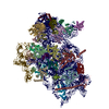
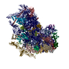
TitleMolecular architecture of 40S translation initiation complexes on the hepatitis C virus IRES.
Journal, issue, pagesEMBO J, Vol. 41, Issue 16, Page e110581, Year 2022
Publish dateAug 16, 2022
AuthorsZuben P Brown / Irina S Abaeva / Swastik De / Christopher U T Hellen / Tatyana V Pestova / Joachim Frank /
PubMed AbstractHepatitis C virus mRNA contains an internal ribosome entry site (IRES) that mediates end-independent translation initiation, requiring a subset of eukaryotic initiation factors (eIFs). Biochemical ...Hepatitis C virus mRNA contains an internal ribosome entry site (IRES) that mediates end-independent translation initiation, requiring a subset of eukaryotic initiation factors (eIFs). Biochemical studies revealed that direct binding of the IRES to the 40S ribosomal subunit places the initiation codon into the P site, where it base pairs with eIF2-bound Met-tRNAiMet forming a 48S initiation complex. Subsequently, eIF5 and eIF5B mediate subunit joining, yielding an elongation-competent 80S ribosome. Initiation can also proceed without eIF2, in which case Met-tRNAiMet is recruited directly by eIF5B. However, the structures of initiation complexes assembled on the HCV IRES, the transitions between different states, and the accompanying conformational changes have remained unknown. To fill these gaps, we now obtained cryo-EM structures of IRES initiation complexes, at resolutions up to 3.5 Å, that cover all major stages from the initial ribosomal association, through eIF2-containing 48S initiation complexes, to eIF5B-containing complexes immediately prior to subunit joining. These structures provide insights into the dynamic network of 40S/IRES contacts, highlight the role of IRES domain II, and reveal conformational changes that occur during the transition from eIF2- to eIF5B-containing 48S complexes and prepare them for subunit joining.
External linksEMBO J / PubMed:35822879 / PubMed Central
MethodsEM (single particle)
Resolution3.1 - 6.2 Å
Structure data

EMDB-25529, PDB-7syi:
Structure of the HCV IRES binding to the 40S ribosomal subunit, closed conformation. Structure 3(delta dII)
Method: EM (single particle) / Resolution: 4.5 Å

EMDB-25530, PDB-7syj:
Structure of the HCV IRES binding to the 40S ribosomal subunit, closed conformation. Structure 4(delta dII)
Method: EM (single particle) / Resolution: 4.8 Å

EMDB-25531, PDB-7syk:
Structure of the HCV IRES binding to the 40S ribosomal subunit, closed conformation. Structure 5(delta dII)
Method: EM (single particle) / Resolution: 4.2 Å

EMDB-25532, PDB-7syl:
Structure of the HCV IRES bound to the 40S ribosomal subunit, closed conformation. Structure 6(delta dII)
Method: EM (single particle) / Resolution: 4.5 Å

EMDB-25535, PDB-7syo:
Structure of the HCV IRES bound to the 40S ribosomal subunit, head open. Structure 9(delta dII)
Method: EM (single particle) / Resolution: 4.6 Å

EMDB-25536, PDB-7syp:
Structure of the wt IRES and 40S ribosome binary complex, open conformation. Structure 10(wt)
Method: EM (single particle) / Resolution: 4.0 Å

EMDB-25537, PDB-7syq:
Structure of the wt IRES and 40S ribosome ternary complex, open conformation. Structure 11(wt)
Method: EM (single particle) / Resolution: 3.8 Å

EMDB-25538, PDB-7syr:
Structure of the wt IRES eIF2-containing 48S initiation complex, closed conformation. Structure 12(wt).
Method: EM (single particle) / Resolution: 3.6 Å

EMDB-25539, PDB-7sys:
Structure of the delta dII IRES eIF2-containing 48S initiation complex, closed conformation. Structure 12(delta dII).
Method: EM (single particle) / Resolution: 3.5 Å

EMDB-25540, PDB-7syt:
Structure of the wt IRES w/o eIF2 48S initiation complex, closed conformation. Structure 13(wt)
Method: EM (single particle) / Resolution: 4.4 Å

EMDB-25541, PDB-7syu:
Structure of the delta dII IRES w/o eIF2 48S initiation complex, closed conformation. Structure 13(delta dII)
Method: EM (single particle) / Resolution: 4.6 Å

EMDB-25542, PDB-7syv:
Structure of the wt IRES eIF5B-containing pre-48S initiation complex, open conformation. Structure 14(wt)
Method: EM (single particle) / Resolution: 3.9 Å

EMDB-25543, PDB-7syw:
Structure of the wt IRES eIF5B-containing 48S initiation complex, closed conformation. Structure 15(wt)
Method: EM (single particle) / Resolution: 3.7 Å

EMDB-25544, PDB-7syx:
Structure of the delta dII IRES eIF5B-containing 48S initiation complex, closed conformation. Structure 15(delta dII)
Method: EM (single particle) / Resolution: 3.7 Å

EMDB-25545: Map of wt IRES eIF2-containing 48S initiation complex with IRES domain II in position 1. Structure 16(wt).
Method: EM (single particle) / Resolution: 3.76 Å

EMDB-25546: Map of wt IRES eIF2-containing 48S initiation complex with IRES domain II in position 2. Structure 17(wt).
Method: EM (single particle) / Resolution: 3.8 Å

EMDB-25547: Map of wt IRES eIF2-containing 48S initiation complex with IRES domain II in position 3. Structure 18(wt)
Method: EM (single particle) / Resolution: 3.7 Å

EMDB-25548: Map of wt IRES eIF2-containing 48S initiation complex with IRES domain II in position 4. Structure 19(wt)
Method: EM (single particle) / Resolution: 3.58 Å

EMDB-25549: Map of wt IRES eIF2-containing 48S initiation complex with IRES domain II in position 5. Structure 20(wt)
Method: EM (single particle) / Resolution: 3.87 Å

EMDB-25550: Map of wt IRES eIF2-containing 48S initiation complex with IRES domain II in position 6. Structure 21(wt)
Method: EM (single particle) / Resolution: 3.76 Å

EMDB-25551: Map of wt IRES eIF5B-containing 48S initiation complex with IRES domain II in position 7. Structure 22(wt)
Method: EM (single particle) / Resolution: 5.0 Å

EMDB-25552: Map of wt IRES eIF5B-containing 48S initiation complex with IRES domain II in position 8. Structure 23(wt)
Method: EM (single particle) / Resolution: 5.4 Å

EMDB-25553: Map of wt IRES eIF5B-containing 48S initiation complex with IRES domain II in position 9. Structure 24(wt)
Method: EM (single particle) / Resolution: 5.2 Å

EMDB-25554: Map of wt IRES eIF5B-containing 48S initiation complex with IRES domain II in position 10. Structure 25(wt)
Method: EM (single particle) / Resolution: 4.9 Å

EMDB-25555: Map of wt IRES eIF5B-containing 48S initiation complex with IRES domain II in position 11. Structure 26(wt)
Method: EM (single particle) / Resolution: 6.0 Å

EMDB-25556: Map of wt IRES eIF5B-containing 48S initiation complex with IRES domain II in position 12. Structure 27(wt)
Method: EM (single particle) / Resolution: 5.3 Å

EMDB-25557: Map of wt IRES eIF5B-containing 48S initiation complex with IRES domain II in position 13. Structure 28(wt)
Method: EM (single particle) / Resolution: 6.2 Å

EMDB-25588: Consensus map of open 40S ribosome from wt IRES eIF2-containing sample
Method: EM (single particle) / Resolution: 3.8 Å

EMDB-25589: Consensus map of open 40S ribosome from wt IRES eIF5B-containing sample
Method: EM (single particle) / Resolution: 4.1 Å

EMDB-25594: Consensus map of open 40S ribosome from wt IRES eIF5B-, and eIF2-containing sample
Method: EM (single particle) / Resolution: 3.3 Å

EMDB-25595: Consensus map of closed 40S ribosome from wt IRES eIF2-containing sample
Method: EM (single particle) / Resolution: 3.1 Å

EMDB-25596: Consensus map of closed 40S ribosome from delta dII IRES eIF2-containing sample
Method: EM (single particle) / Resolution: 3.3 Å

EMDB-25597: Consensus map of open 40S ribosome from wt IRES eIF5B-containing sample
Method: EM (single particle) / Resolution: 3.6 Å

EMDB-25598: Consensus map of closed 40S ribosome from wt IRES eIF5B-containing sample
Method: EM (single particle) / Resolution: 3.4 Å

EMDB-25599: Consensus map of closed 40S ribosome from delta dII IRES eIF5B-containing sample
Method: EM (single particle) / Resolution: 3.4 Å

EMDB-25602: Map of the wt IRES eIF5B-containing 48S initiation complex, closed conformation
Method: EM (single particle) / Resolution: 3.9 Å

EMDB-25603: Map of the wt IRES eIF5B-containing pre-48S initiation complex, open conformation
Method: EM (single particle) / Resolution: 4.2 Å

EMDB-25604: Map of the wt IRES eIF2-containing 48S initiation complex, closed conformation
Method: EM (single particle) / Resolution: 3.6 Å

EMDB-25620: Map of the delta IRES eIF2-containing 48S initiation complex w/o eIF2, closed conformation
Method: EM (single particle) / Resolution: 3.55 Å

Chemicals

ChemComp-ZN:
Unknown entry

ChemComp-GTP:
GUANOSINE-5'-TRIPHOSPHATE / GTP, energy-carrying molecule*YM / Guanosine triphosphate

ChemComp-MG:
Unknown entry

ChemComp-NA:
Unknown entry

Source
  • rabbit (rabbit)
  • hepacivirus c
  • hepatitis c virus genotype 1a
  • oryctolagus cuniculus (rabbit)
  • hepatitis c virus (isolate 1)
  • homo sapiens (human)
KeywordsRIBOSOME / HCV / IRES / 40S

+
About Yorodumi Papers

-
News

-
Feb 9, 2022. New format data for meta-information of EMDB entries

New format data for meta-information of EMDB entries

  • Version 3 of the EMDB header file is now the official format.
  • The previous official version 1.9 will be removed from the archive.

Related info.:EMDB header

External links:wwPDB to switch to version 3 of the EMDB data model

-
Aug 12, 2020. Covid-19 info

Covid-19 info

URL: https://pdbj.org/emnavi/covid19.php

New page: Covid-19 featured information page in EM Navigator.

Related info.:Covid-19 info / Mar 5, 2020. Novel coronavirus structure data

+
Mar 5, 2020. Novel coronavirus structure data

Novel coronavirus structure data

Related info.:Yorodumi Speices / Aug 12, 2020. Covid-19 info

External links:COVID-19 featured content - PDBj / Molecule of the Month (242):Coronavirus Proteases

+
Jan 31, 2019. EMDB accession codes are about to change! (news from PDBe EMDB page)

EMDB accession codes are about to change! (news from PDBe EMDB page)

  • The allocation of 4 digits for EMDB accession codes will soon come to an end. Whilst these codes will remain in use, new EMDB accession codes will include an additional digit and will expand incrementally as the available range of codes is exhausted. The current 4-digit format prefixed with “EMD-” (i.e. EMD-XXXX) will advance to a 5-digit format (i.e. EMD-XXXXX), and so on. It is currently estimated that the 4-digit codes will be depleted around Spring 2019, at which point the 5-digit format will come into force.
  • The EM Navigator/Yorodumi systems omit the EMD- prefix.

Related info.:Q: What is EMD? / ID/Accession-code notation in Yorodumi/EM Navigator

External links:EMDB Accession Codes are Changing Soon! / Contact to PDBj

+
Jul 12, 2017. Major update of PDB

Major update of PDB

  • wwPDB released updated PDB data conforming to the new PDBx/mmCIF dictionary.
  • This is a major update changing the version number from 4 to 5, and with Remediation, in which all the entries are updated.
  • In this update, many items about electron microscopy experimental information are reorganized (e.g. em_software).
  • Now, EM Navigator and Yorodumi are based on the updated data.

External links:wwPDB Remediation / Enriched Model Files Conforming to OneDep Data Standards Now Available in the PDB FTP Archive

-
Yorodumi Papers

Database of articles cited by EMDB/PDB/SASBDB data

  • Database of articles cited by EMDB, PDB, and SASBDB entries
  • Using PubMed data

Related info.:EMDB / PDB / SASBDB / Yorodumi / EMN Papers / Changes in new EM Navigator and Yorodumi

Read more