[English] 日本語
Yorodumi
- PDB-7syq: Structure of the wt IRES and 40S ribosome ternary complex, open c... -

+
Open data


ID or keywords:

Loading...

-
Basic information

Entry
Database: PDB / ID: 7syq
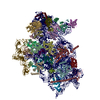
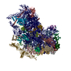
TitleStructure of the wt IRES and 40S ribosome ternary complex, open conformation. Structure 11(wt)
Components
  • 18S rRNA18S ribosomal RNA
  • Eukaryotic translation initiation factor 1A, X-chromosomal
  • HCV IRESHepatitis C virus internal ribosome entry site
  • Receptor for Activated C Kinase 1 (RACK1)
  • eL41
  • eS1
  • eS10
  • eS12
  • eS17
  • eS19
  • eS21
  • eS24
  • eS25
  • eS26
  • eS27
  • eS28
  • eS30
  • eS31
  • eS4 (S4 X isoform)
  • eS6
  • eS7
  • eS8
  • uS10
  • uS11
  • uS12
  • uS13
  • uS14
  • uS15
  • uS17
  • uS19
  • uS2 (SA)
  • uS3
  • uS4
  • uS5
  • uS7
  • uS8
  • uS9
KeywordsRIBOSOME / HCV / IRES / 40S
Function / homology
Function and homology information


multi-eIF complex / eukaryotic 43S preinitiation complex / translation factor activity, RNA binding / eukaryotic 48S preinitiation complex / ribosomal subunit / Formation of the ternary complex, and subsequently, the 43S complex / Ribosomal scanning and start codon recognition / Translation initiation complex formation / Formation of a pool of free 40S subunits / GTP hydrolysis and joining of the 60S ribosomal subunit ...multi-eIF complex / eukaryotic 43S preinitiation complex / translation factor activity, RNA binding / eukaryotic 48S preinitiation complex / ribosomal subunit / Formation of the ternary complex, and subsequently, the 43S complex / Ribosomal scanning and start codon recognition / Translation initiation complex formation / Formation of a pool of free 40S subunits / GTP hydrolysis and joining of the 60S ribosomal subunit / L13a-mediated translational silencing of Ceruloplasmin expression / translational initiation / translation initiation factor activity / ribosome assembly / DNA-(apurinic or apyrimidinic site) lyase / tRNA binding / structural constituent of ribosome / translation / synapse / RNA binding / cytosol / cytoplasm
Similarity search - Function
Translation initiation factor 1A (eIF-1A), conserved site / Eukaryotic initiation factor 1A signature. / eukaryotic translation initiation factor 1A / Translation initiation factor 1A (eIF-1A) / RNA-binding domain, S1, IF1 type / Translation initiation factor 1A / IF-1 / S1 domain IF1 type profile. / Ribosomal protein S26e signature. / Ribosomal protein S21e signature. / Ribosomal protein S12e signature. ...Translation initiation factor 1A (eIF-1A), conserved site / Eukaryotic initiation factor 1A signature. / eukaryotic translation initiation factor 1A / Translation initiation factor 1A (eIF-1A) / RNA-binding domain, S1, IF1 type / Translation initiation factor 1A / IF-1 / S1 domain IF1 type profile. / Ribosomal protein S26e signature. / Ribosomal protein S21e signature. / Ribosomal protein S12e signature. / Ribosomal protein S19e signature. / 40S Ribosomal protein S10 / Plectin/S10, N-terminal / Plectin/S10 domain / Ribosomal protein S17e signature. / Ribosomal protein S7e signature. / Ribosomal protein S3Ae signature. / Ribosomal protein S27e signature. / Ribosomal protein S4e signature. / Ribosomal protein S8e signature. / Ribosomal S24e conserved site / Ribosomal protein S24e signature. / Ribosomal protein S24e / Ribosomal protein S24e / Ribosomal protein S6e signature. / Ribosomal protein S28e signature. / Ubiquitin domain signature. / Ribosomal protein S2 signature 2. / Ribosomal protein S3 signature. / Ribosomal protein S10 signature. / Ribosomal protein S14 signature. / Ribosomal protein S2 signature 1. / Type-2 KH domain profile. / Ribosomal protein S19 signature. / Ribosomal protein S7 signature. / Ribosomal protein S17 signature. / Ribosomal protein S13 signature. / Ribosomal protein S13 family profile. / Ribosomal protein S8 signature. / Ribosomal protein S4 signature. / Ribosomal protein S15 signature. / S4 RNA-binding domain profile. / Ribosomal protein S11 signature. / Ribosomal protein S9 signature. / Ubiquitin domain profile. / Ribosomal protein S12 signature. / Ribosomal protein L23/L15e core domain superfamily / Trp-Asp (WD) repeats signature. / Trp-Asp (WD) repeats profile. / Trp-Asp (WD) repeats circular profile. / Winged helix-like DNA-binding domain superfamily / Nucleic acid-binding, OB-fold
Similarity search - Domain/homology
: / RNA / RNA (> 10) / RNA (> 100) / RNA (> 1000) / Small ribosomal subunit protein eS32 / Small ribosomal subunit protein uS4 / Small ribosomal subunit protein eS12 / Small ribosomal subunit protein uS9 / Small ribosomal subunit protein uS10 ...: / RNA / RNA (> 10) / RNA (> 100) / RNA (> 1000) / Small ribosomal subunit protein eS32 / Small ribosomal subunit protein uS4 / Small ribosomal subunit protein eS12 / Small ribosomal subunit protein uS9 / Small ribosomal subunit protein uS10 / Small ribosomal subunit protein RACK1 / Ubiquitin-ribosomal protein eS31 fusion protein / Small ribosomal subunit protein uS15 / Small ribosomal subunit protein eS1 / Small ribosomal subunit protein eS7 / Small ribosomal subunit protein uS12 / 40S ribosomal protein S24 / Ubiquitin-like FUBI-ribosomal protein eS30 fusion protein / Small ribosomal subunit protein eS25 / Small ribosomal subunit protein eS26 / Small ribosomal subunit protein uS7 / Small ribosomal subunit protein uS8 / Small ribosomal subunit protein eS28 / Small ribosomal subunit protein eS8 / Small ribosomal subunit protein eS4 / Small ribosomal subunit protein uS2 / Small ribosomal subunit protein eS6 / Small ribosomal subunit protein eS21 / Small ribosomal subunit protein eS19 / Small ribosomal subunit protein uS3 / Small ribosomal subunit protein uS13 / Plectin/eS10 N-terminal domain-containing protein / Small ribosomal subunit protein uS17 / Small ribosomal subunit protein eS17 / Small ribosomal subunit protein eS27 / Small ribosomal subunit protein uS19 / Small ribosomal subunit protein uS11 / Small ribosomal subunit protein uS14 / Eukaryotic translation initiation factor 1A, X-chromosomal
Similarity search - Component
Biological speciesHomo sapiens (human)
Hepatitis C virus
Oryctolagus cuniculus (rabbit)
MethodELECTRON MICROSCOPY / single particle reconstruction / cryo EM / Resolution: 3.8 Å
AuthorsBrown, Z.P. / Abaeva, I.S. / De, S. / Hellen, C.U.T. / Pestova, T.V. / Frank, J.
Funding support United States, 4items
OrganizationGrant numberCountry
National Institutes of Health/National Institute of General Medical Sciences (NIH/NIGMS)R35 GM139453 United States
National Institutes of Health/National Institute of General Medical Sciences (NIH/NIGMS)R01 GM55440 United States
National Institutes of Health/National Institute of General Medical Sciences (NIH/NIGMS)1R35GM122602 United States
National Institutes of Health/National Institute Of Allergy and Infectious Diseases (NIH/NIAID)1R01AI123406 United States
CitationJournal: EMBO J / Year: 2022
Title: Molecular architecture of 40S translation initiation complexes on the hepatitis C virus IRES.
Authors: Zuben P Brown / Irina S Abaeva / Swastik De / Christopher U T Hellen / Tatyana V Pestova / Joachim Frank /
Abstract: Hepatitis C virus mRNA contains an internal ribosome entry site (IRES) that mediates end-independent translation initiation, requiring a subset of eukaryotic initiation factors (eIFs). Biochemical ...Hepatitis C virus mRNA contains an internal ribosome entry site (IRES) that mediates end-independent translation initiation, requiring a subset of eukaryotic initiation factors (eIFs). Biochemical studies revealed that direct binding of the IRES to the 40S ribosomal subunit places the initiation codon into the P site, where it base pairs with eIF2-bound Met-tRNAiMet forming a 48S initiation complex. Subsequently, eIF5 and eIF5B mediate subunit joining, yielding an elongation-competent 80S ribosome. Initiation can also proceed without eIF2, in which case Met-tRNAiMet is recruited directly by eIF5B. However, the structures of initiation complexes assembled on the HCV IRES, the transitions between different states, and the accompanying conformational changes have remained unknown. To fill these gaps, we now obtained cryo-EM structures of IRES initiation complexes, at resolutions up to 3.5 Å, that cover all major stages from the initial ribosomal association, through eIF2-containing 48S initiation complexes, to eIF5B-containing complexes immediately prior to subunit joining. These structures provide insights into the dynamic network of 40S/IRES contacts, highlight the role of IRES domain II, and reveal conformational changes that occur during the transition from eIF2- to eIF5B-containing 48S complexes and prepare them for subunit joining.
History
DepositionNov 25, 2021Deposition site: RCSB / Processing site: RCSB
Revision 1.0Jul 27, 2022Provider: repository / Type: Initial release
Revision 1.1Aug 24, 2022Group: Database references / Category: citation / Item: _citation.journal_volume

-
Structure visualization

Structure viewerMolecule:
MolmilJmol/JSmol

Downloads & links

-
Assembly

Deposited unit
2: 18S rRNA
A: Eukaryotic translation initiation factor 1A, X-chromosomal
B: uS2 (SA)
C: eS1
D: uS5
E: uS3
F: eS4 (S4 X isoform)
G: uS7
H: eS6
I: eS7
J: eS8
K: uS4
L: eS10
M: uS17
N: eS12
O: uS15
P: uS11
Q: uS19
R: uS9
S: eS17
T: uS13
U: eS19
V: uS10
W: eS21
X: uS8
Y: uS12
Z: eS24
a: eS25
b: eS26
c: eS27
d: eS28
e: uS14
f: eS30
g: eS31
h: Receptor for Activated C Kinase 1 (RACK1)
n: eL41
z: HCV IRES
hetero molecules


Theoretical massNumber of molelcules
Total (without water)1,406,27239
Polymers1,406,14137
Non-polymers1312
Water0
1


  • Idetical with deposited unit
  • defined by author
  • Evidence: electron microscopy
TypeNameSymmetry operationNumber
identity operation1_5551

-
Components

-
RNA chain , 2 types, 2 molecules 2z

#1: RNA chain 18S rRNA / 18S ribosomal RNA


Mass: 603100.938 Da / Num. of mol.: 1 / Source method: isolated from a natural source / Source: (natural) Oryctolagus cuniculus (rabbit)
#37: RNA chain HCV IRES / Hepatitis C virus internal ribosome entry site


Mass: 128746.109 Da / Num. of mol.: 1
Source method: isolated from a genetically manipulated source
Source: (gene. exp.) Hepatitis C virus (isolate 1) / Production host: Escherichia coli (E. coli) / References: GenBank: 149384897

+
Protein , 34 types, 34 molecules ABCDEFGHIJKLMNOPQRSTUVWXYZabcd...

#2: Protein Eukaryotic translation initiation factor 1A, X-chromosomal / eIF-1A X isoform / Eukaryotic translation initiation factor 4C / eIF-4C


Mass: 16488.449 Da / Num. of mol.: 1
Source method: isolated from a genetically manipulated source
Source: (gene. exp.) Homo sapiens (human) / Gene: EIF1AX, EIF1A, EIF4C / Production host: Escherichia coli (E. coli) / References: UniProt: P47813
#3: Protein uS2 (SA) / 40S ribosomal protein SA / 37 kDa laminin receptor precursor / 37LRP / 37/67 kDa laminin receptor / ...40S ribosomal protein SA / 37 kDa laminin receptor precursor / 37LRP / 37/67 kDa laminin receptor / LRP/LR / 67 kDa laminin receptor / 67LR / Laminin receptor 1 / LamR / Laminin-binding protein precursor p40 / LBP/p40


Mass: 32958.016 Da / Num. of mol.: 1 / Mutation: T114A / Source method: isolated from a natural source / Source: (natural) Oryctolagus cuniculus (rabbit) / References: UniProt: G1TLT8
#4: Protein eS1 / 40S ribosomal protein S3a


Mass: 30002.061 Da / Num. of mol.: 1 / Source method: isolated from a natural source / Source: (natural) Oryctolagus cuniculus (rabbit) / References: UniProt: G1SS70
#5: Protein uS5


Mass: 24441.846 Da / Num. of mol.: 1 / Source method: isolated from a natural source / Source: (natural) Oryctolagus cuniculus (rabbit)
#6: Protein uS3 / Ribosomal protein S3


Mass: 31146.607 Da / Num. of mol.: 1 / Source method: isolated from a natural source / Source: (natural) Oryctolagus cuniculus (rabbit) / References: UniProt: G1TNM3
#7: Protein eS4 (S4 X isoform) / 40S ribosomal protein S4


Mass: 29654.869 Da / Num. of mol.: 1 / Source method: isolated from a natural source / Source: (natural) Oryctolagus cuniculus (rabbit) / References: UniProt: G1TK17
#8: Protein uS7 / 40S ribosomal protein S5


Mass: 22913.453 Da / Num. of mol.: 1 / Source method: isolated from a natural source / Source: (natural) Oryctolagus cuniculus (rabbit) / References: UniProt: G1TFM5
#9: Protein eS6 / 40S ribosomal protein S6


Mass: 28751.906 Da / Num. of mol.: 1 / Source method: isolated from a natural source / Source: (natural) Oryctolagus cuniculus (rabbit) / References: UniProt: G1TM55
#10: Protein eS7 / 40S ribosomal protein S7


Mass: 47356.172 Da / Num. of mol.: 1 / Source method: isolated from a natural source / Source: (natural) Oryctolagus cuniculus (rabbit) / References: UniProt: G1SVB0
#11: Protein eS8 / 40S ribosomal protein S8


Mass: 24263.387 Da / Num. of mol.: 1 / Source method: isolated from a natural source / Source: (natural) Oryctolagus cuniculus (rabbit) / References: UniProt: G1TJW1
#12: Protein uS4 / Ribosomal protein S9


Mass: 22641.564 Da / Num. of mol.: 1 / Source method: isolated from a natural source / Source: (natural) Oryctolagus cuniculus (rabbit) / References: UniProt: B7NZS8
#13: Protein eS10


Mass: 17156.865 Da / Num. of mol.: 1 / Source method: isolated from a natural source / Source: (natural) Oryctolagus cuniculus (rabbit) / References: UniProt: G1TPV3
#14: Protein uS17 / 40S ribosomal protein S11


Mass: 18468.826 Da / Num. of mol.: 1 / Source method: isolated from a natural source / Source: (natural) Oryctolagus cuniculus (rabbit) / References: UniProt: G1TRM4
#15: Protein eS12 / 40S ribosomal protein S12


Mass: 14538.987 Da / Num. of mol.: 1 / Source method: isolated from a natural source / Source: (natural) Oryctolagus cuniculus (rabbit) / References: UniProt: G1SFR8
#16: Protein uS15 / Ribosomal protein S13


Mass: 17259.389 Da / Num. of mol.: 1 / Source method: isolated from a natural source / Source: (natural) Oryctolagus cuniculus (rabbit) / References: UniProt: G1SP51
#17: Protein uS11


Mass: 18133.984 Da / Num. of mol.: 1 / Source method: isolated from a natural source / Source: (natural) Oryctolagus cuniculus (rabbit) / References: UniProt: G1U472
#18: Protein uS19 / 40S ribosomal protein S15


Mass: 17049.182 Da / Num. of mol.: 1 / Source method: isolated from a natural source / Source: (natural) Oryctolagus cuniculus (rabbit) / References: UniProt: G1U0Q2
#19: Protein uS9 / Ribosomal protein S16


Mass: 19213.465 Da / Num. of mol.: 1 / Source method: isolated from a natural source / Source: (natural) Oryctolagus cuniculus (rabbit) / References: UniProt: G1SGX4
#20: Protein eS17 / 40S ribosomal protein S17


Mass: 15552.119 Da / Num. of mol.: 1 / Source method: isolated from a natural source / Source: (natural) Oryctolagus cuniculus (rabbit) / References: UniProt: G1TU13
#21: Protein uS13 / 40S ribosomal protein S18


Mass: 17759.777 Da / Num. of mol.: 1 / Source method: isolated from a natural source / Source: (natural) Oryctolagus cuniculus (rabbit) / References: UniProt: G1TPG3
#22: Protein eS19 / 40S ribosomal protein S19


Mass: 16235.796 Da / Num. of mol.: 1 / Source method: isolated from a natural source / Source: (natural) Oryctolagus cuniculus (rabbit) / References: UniProt: G1TN62
#23: Protein uS10 / 40S ribosomal protein S20


Mass: 13398.763 Da / Num. of mol.: 1 / Source method: isolated from a natural source / Source: (natural) Oryctolagus cuniculus (rabbit) / References: UniProt: G1SIZ2
#24: Protein eS21 / 40S ribosomal protein S21


Mass: 9124.389 Da / Num. of mol.: 1 / Source method: isolated from a natural source / Source: (natural) Oryctolagus cuniculus (rabbit) / References: UniProt: G1TM82
#25: Protein uS8 / 40S Ribosomal protein S15a


Mass: 14865.555 Da / Num. of mol.: 1 / Source method: isolated from a natural source / Source: (natural) Oryctolagus cuniculus (rabbit) / References: UniProt: G1TG89
#26: Protein uS12 / 40S ribosomal protein S23


Mass: 15844.666 Da / Num. of mol.: 1 / Source method: isolated from a natural source / Source: (natural) Oryctolagus cuniculus (rabbit) / References: UniProt: G1SZ47
#27: Protein eS24 / 40S ribosomal protein S24


Mass: 15237.104 Da / Num. of mol.: 1 / Source method: isolated from a natural source / Source: (natural) Oryctolagus cuniculus (rabbit) / References: UniProt: G1T3D8
#28: Protein eS25 / 40S ribosomal protein S25


Mass: 13645.028 Da / Num. of mol.: 1 / Source method: isolated from a natural source / Source: (natural) Oryctolagus cuniculus (rabbit) / References: UniProt: G1TDB3
#29: Protein eS26 / 40S ribosomal protein S26


Mass: 11645.794 Da / Num. of mol.: 1 / Source method: isolated from a natural source / Source: (natural) Oryctolagus cuniculus (rabbit) / References: UniProt: G1TFE8
#30: Protein eS27 / 40S ribosomal protein S27


Mass: 9480.186 Da / Num. of mol.: 1 / Source method: isolated from a natural source / Source: (natural) Oryctolagus cuniculus (rabbit) / References: UniProt: G1TZ76
#31: Protein eS28 / 40S Ribosomal protein S28


Mass: 7855.052 Da / Num. of mol.: 1 / Source method: isolated from a natural source / Source: (natural) Oryctolagus cuniculus (rabbit) / References: UniProt: G1TIB4
#32: Protein uS14 / 40S ribosomal protein S29


Mass: 6690.821 Da / Num. of mol.: 1 / Source method: isolated from a natural source / Source: (natural) Oryctolagus cuniculus (rabbit) / References: UniProt: G1U7M4
#33: Protein eS30 / 40S ribosomal protein S30


Mass: 14498.884 Da / Num. of mol.: 1 / Source method: isolated from a natural source / Source: (natural) Oryctolagus cuniculus (rabbit) / References: UniProt: G1T8A2
#34: Protein eS31 / 40S ribosomal protein S27a / Ubiquitin carboxyl extension protein 80 / Ubiquitin-40S ribosomal protein S27a


Mass: 21431.742 Da / Num. of mol.: 1 / Source method: isolated from a natural source / Source: (natural) Oryctolagus cuniculus (rabbit) / References: UniProt: G1SK22
#35: Protein Receptor for Activated C Kinase 1 (RACK1)


Mass: 35115.652 Da / Num. of mol.: 1 / Source method: isolated from a natural source / Source: (natural) Oryctolagus cuniculus (rabbit) / References: UniProt: G1SJB4

-
Protein/peptide / Non-polymers , 2 types, 3 molecules n

#36: Protein/peptide eL41 / 60s ribosomal protein l41


Mass: 3473.451 Da / Num. of mol.: 1 / Source method: isolated from a natural source / Source: (natural) Oryctolagus cuniculus (rabbit) / References: UniProt: A0A087WNH4
#38: Chemical ChemComp-ZN / ZINC ION


Mass: 65.409 Da / Num. of mol.: 2 / Source method: obtained synthetically / Formula: Zn

-
Details

Has ligand of interestN

-
Experimental details

-
Experiment

ExperimentMethod: ELECTRON MICROSCOPY
EM experimentAggregation state: PARTICLE / 3D reconstruction method: single particle reconstruction

-
Sample preparation

ComponentName: 40S ribosomal small subunit with HCV IRES / Type: RIBOSOME / Entity ID: #1-#37 / Source: MULTIPLE SOURCES
Molecular weightValue: 2 MDa / Experimental value: NO
Buffer solutionpH: 7.5
SpecimenConc.: 7.5E-5 mg/ml / Embedding applied: NO / Shadowing applied: NO / Staining applied: NO / Vitrification applied: YES
Specimen supportDetails: H2/O2 mixture for 25 seconds at 25W power / Grid material: GOLD / Grid mesh size: 300 divisions/in. / Grid type: Quantifoil R0.6/1
VitrificationInstrument: FEI VITROBOT MARK IV / Cryogen name: ETHANE-PROPANE / Humidity: 100 % / Chamber temperature: 277.15 K / Details: 4 second blot time, force 3

-
Electron microscopy imaging

Experimental equipment
Model: Tecnai F30 / Image courtesy: FEI Company
MicroscopyModel: FEI TECNAI F30
Electron gunElectron source: FIELD EMISSION GUN / Accelerating voltage: 300 kV / Illumination mode: FLOOD BEAM
Electron lensMode: BRIGHT FIELDBright-field microscopy / Nominal magnification: 56000 X / Nominal defocus max: 2000 nm / Nominal defocus min: 1000 nm / Cs: 2.26 mm / Alignment procedure: COMA FREE
Specimen holderCryogen: NITROGEN
Image recordingAverage exposure time: 4 sec. / Electron dose: 70.9 e/Å2 / Film or detector model: GATAN K3 (6k x 4k)

-
Processing

SoftwareName: PHENIX / Version: 1.19.1_4122: / Classification: refinement
EM software
IDNameVersionCategory
2Leginonimage acquisition
4CTFFINDCTF correction
7UCSF Chimera1.15model fitting
9RELION3.1initial Euler assignment
12RELION3.13D reconstruction
CTF correctionType: PHASE FLIPPING AND AMPLITUDE CORRECTION
SymmetryPoint symmetry: C1 (asymmetric)
3D reconstructionResolution: 3.8 Å / Resolution method: FSC 0.143 CUT-OFF / Num. of particles: 204782 / Algorithm: BACK PROJECTION / Symmetry type: POINT
Refine LS restraints
Refine-IDTypeDev idealNumber
ELECTRON MICROSCOPYf_bond_d0.00386999
ELECTRON MICROSCOPYf_angle_d0.773126768
ELECTRON MICROSCOPYf_dihedral_angle_d15.01328848
ELECTRON MICROSCOPYf_chiral_restr0.0415742
ELECTRON MICROSCOPYf_plane_restr0.0078870

+
About Yorodumi

-
News

-
Feb 9, 2022. New format data for meta-information of EMDB entries

New format data for meta-information of EMDB entries

  • Version 3 of the EMDB header file is now the official format.
  • The previous official version 1.9 will be removed from the archive.

Related info.:EMDB header

External links:wwPDB to switch to version 3 of the EMDB data model

-
Aug 12, 2020. Covid-19 info

Covid-19 info

URL: https://pdbj.org/emnavi/covid19.php

New page: Covid-19 featured information page in EM Navigator.

Related info.:Covid-19 info / Mar 5, 2020. Novel coronavirus structure data

+
Mar 5, 2020. Novel coronavirus structure data

Novel coronavirus structure data

Related info.:Yorodumi Speices / Aug 12, 2020. Covid-19 info

External links:COVID-19 featured content - PDBj / Molecule of the Month (242):Coronavirus Proteases

+
Jan 31, 2019. EMDB accession codes are about to change! (news from PDBe EMDB page)

EMDB accession codes are about to change! (news from PDBe EMDB page)

  • The allocation of 4 digits for EMDB accession codes will soon come to an end. Whilst these codes will remain in use, new EMDB accession codes will include an additional digit and will expand incrementally as the available range of codes is exhausted. The current 4-digit format prefixed with “EMD-” (i.e. EMD-XXXX) will advance to a 5-digit format (i.e. EMD-XXXXX), and so on. It is currently estimated that the 4-digit codes will be depleted around Spring 2019, at which point the 5-digit format will come into force.
  • The EM Navigator/Yorodumi systems omit the EMD- prefix.

Related info.:Q: What is EMD? / ID/Accession-code notation in Yorodumi/EM Navigator

External links:EMDB Accession Codes are Changing Soon! / Contact to PDBj

+
Jul 12, 2017. Major update of PDB

Major update of PDB

  • wwPDB released updated PDB data conforming to the new PDBx/mmCIF dictionary.
  • This is a major update changing the version number from 4 to 5, and with Remediation, in which all the entries are updated.
  • In this update, many items about electron microscopy experimental information are reorganized (e.g. em_software).
  • Now, EM Navigator and Yorodumi are based on the updated data.

External links:wwPDB Remediation / Enriched Model Files Conforming to OneDep Data Standards Now Available in the PDB FTP Archive

-
Yorodumi

Thousand views of thousand structures

  • Yorodumi is a browser for structure data from EMDB, PDB, SASBDB, etc.
  • This page is also the successor to EM Navigator detail page, and also detail information page/front-end page for Omokage search.
  • The word "yorodu" (or yorozu) is an old Japanese word meaning "ten thousand". "mi" (miru) is to see.

Related info.:EMDB / PDB / SASBDB / Comparison of 3 databanks / Yorodumi Search / Aug 31, 2016. New EM Navigator & Yorodumi / Yorodumi Papers / Jmol/JSmol / Function and homology information / Changes in new EM Navigator and Yorodumi

Read more