 Movie
Movie Controller
 Structure viewers
 About Yorodumi Papers


+Search query
-Structure paper
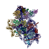
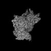
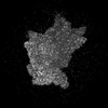
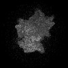
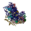
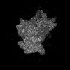
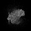
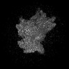
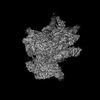
| Title | Molecular architecture of 40S translation initiation complexes on the hepatitis C virus IRES. |
|---|---|
| Journal, issue, pages | EMBO J, Vol. 41, Issue 16, Page e110581, Year 2022 |
| Publish date | Aug 16, 2022 |
 Authors Authors | Zuben P Brown / Irina S Abaeva / Swastik De / Christopher U T Hellen / Tatyana V Pestova / Joachim Frank /  |
| PubMed Abstract | Hepatitis C virus mRNA contains an internal ribosome entry site (IRES) that mediates end-independent translation initiation, requiring a subset of eukaryotic initiation factors (eIFs). Biochemical ...Hepatitis C virus mRNA contains an internal ribosome entry site (IRES) that mediates end-independent translation initiation, requiring a subset of eukaryotic initiation factors (eIFs). Biochemical studies revealed that direct binding of the IRES to the 40S ribosomal subunit places the initiation codon into the P site, where it base pairs with eIF2-bound Met-tRNAiMet forming a 48S initiation complex. Subsequently, eIF5 and eIF5B mediate subunit joining, yielding an elongation-competent 80S ribosome. Initiation can also proceed without eIF2, in which case Met-tRNAiMet is recruited directly by eIF5B. However, the structures of initiation complexes assembled on the HCV IRES, the transitions between different states, and the accompanying conformational changes have remained unknown. To fill these gaps, we now obtained cryo-EM structures of IRES initiation complexes, at resolutions up to 3.5 Å, that cover all major stages from the initial ribosomal association, through eIF2-containing 48S initiation complexes, to eIF5B-containing complexes immediately prior to subunit joining. These structures provide insights into the dynamic network of 40S/IRES contacts, highlight the role of IRES domain II, and reveal conformational changes that occur during the transition from eIF2- to eIF5B-containing 48S complexes and prepare them for subunit joining. |
 External links External links | EMBO J / PubMed:35822879 / PubMed Central |
| Methods | EM (single particle) |
| Resolution | 3.1 - 6.2 Å |
| Structure data | EMDB-25529, PDB-7syi: EMDB-25530, PDB-7syj: EMDB-25531, PDB-7syk: EMDB-25532, PDB-7syl: EMDB-25535, PDB-7syo: EMDB-25536, PDB-7syp: EMDB-25537, PDB-7syq: EMDB-25538, PDB-7syr: EMDB-25539, PDB-7sys: EMDB-25540, PDB-7syt: EMDB-25541, PDB-7syu: EMDB-25542, PDB-7syv: EMDB-25543, PDB-7syw: EMDB-25544, PDB-7syx:  EMDB-25545: Map of wt IRES eIF2-containing 48S initiation complex with IRES domain II in position 1. Structure 16(wt).  EMDB-25546: Map of wt IRES eIF2-containing 48S initiation complex with IRES domain II in position 2. Structure 17(wt).  EMDB-25547: Map of wt IRES eIF2-containing 48S initiation complex with IRES domain II in position 3. Structure 18(wt)  EMDB-25548: Map of wt IRES eIF2-containing 48S initiation complex with IRES domain II in position 4. Structure 19(wt)  EMDB-25549: Map of wt IRES eIF2-containing 48S initiation complex with IRES domain II in position 5. Structure 20(wt)  EMDB-25550: Map of wt IRES eIF2-containing 48S initiation complex with IRES domain II in position 6. Structure 21(wt)  EMDB-25551: Map of wt IRES eIF5B-containing 48S initiation complex with IRES domain II in position 7. Structure 22(wt)  EMDB-25552: Map of wt IRES eIF5B-containing 48S initiation complex with IRES domain II in position 8. Structure 23(wt)  EMDB-25553: Map of wt IRES eIF5B-containing 48S initiation complex with IRES domain II in position 9. Structure 24(wt)  EMDB-25554: Map of wt IRES eIF5B-containing 48S initiation complex with IRES domain II in position 10. Structure 25(wt)  EMDB-25555: Map of wt IRES eIF5B-containing 48S initiation complex with IRES domain II in position 11. Structure 26(wt)  EMDB-25556: Map of wt IRES eIF5B-containing 48S initiation complex with IRES domain II in position 12. Structure 27(wt)  EMDB-25557: Map of wt IRES eIF5B-containing 48S initiation complex with IRES domain II in position 13. Structure 28(wt)  EMDB-25588: Consensus map of open 40S ribosome from wt IRES eIF2-containing sample  EMDB-25589: Consensus map of open 40S ribosome from wt IRES eIF5B-containing sample  EMDB-25594: Consensus map of open 40S ribosome from wt IRES eIF5B-, and eIF2-containing sample  EMDB-25595: Consensus map of closed 40S ribosome from wt IRES eIF2-containing sample  EMDB-25596: Consensus map of closed 40S ribosome from delta dII IRES eIF2-containing sample  EMDB-25597: Consensus map of open 40S ribosome from wt IRES eIF5B-containing sample  EMDB-25598: Consensus map of closed 40S ribosome from wt IRES eIF5B-containing sample  EMDB-25599: Consensus map of closed 40S ribosome from delta dII IRES eIF5B-containing sample  EMDB-25602: Map of the wt IRES eIF5B-containing 48S initiation complex, closed conformation  EMDB-25603: Map of the wt IRES eIF5B-containing pre-48S initiation complex, open conformation  EMDB-25604: Map of the wt IRES eIF2-containing 48S initiation complex, closed conformation  EMDB-25620: Map of the delta IRES eIF2-containing 48S initiation complex w/o eIF2, closed conformation |
| Chemicals |  ChemComp-ZN:  ChemComp-GTP:  ChemComp-MG:  ChemComp-NA: |
| Source |
|
 Keywords Keywords | RIBOSOME / HCV / IRES / 40S |


 hepacivirus c
 homo sapiens (human)