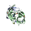
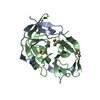
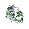
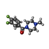
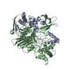
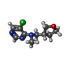
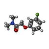
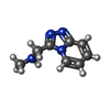
PDB-7h1h: PanDDA analysis group deposition -- Crystal Structure of ZIKV NS2B-NS3 protease in complex with Z1201620232 Method: X-RAY DIFFRACTION / Resolution: 1.684 Å
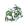
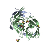
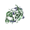
PDB-7h1i: PanDDA analysis group deposition -- Crystal Structure of ZIKV NS2B-NS3 protease in complex with Z336080990 Method: X-RAY DIFFRACTION / Resolution: 1.414 Å
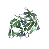
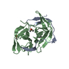
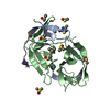
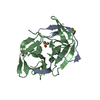
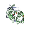
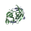
PDB-7h1j: PanDDA analysis group deposition -- Crystal Structure of ZIKV NS2B-NS3 protease in complex with Z68299550 Method: X-RAY DIFFRACTION / Resolution: 1.55 Å
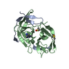
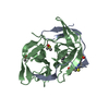
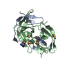
PDB-7h1k: PanDDA analysis group deposition -- Crystal Structure of ZIKV NS2B-NS3 protease in complex with Z57122377 Method: X-RAY DIFFRACTION / Resolution: 1.505 Å
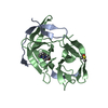
PDB-7h1l: PanDDA analysis group deposition -- Crystal Structure of ZIKV NS2B-NS3 protease in complex with POB0087 Method: X-RAY DIFFRACTION / Resolution: 1.535 Å
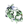
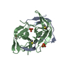
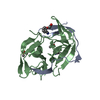
PDB-7h1m: PanDDA analysis group deposition -- Crystal Structure of ZIKV NS2B-NS3 protease in complex with POB0075 Method: X-RAY DIFFRACTION / Resolution: 1.609 Å
PDB-7h1n: PanDDA analysis group deposition -- Crystal Structure of ZIKV NS2B-NS3 protease in complex with POB0120 Method: X-RAY DIFFRACTION / Resolution: 1.407 Å
PDB-7h1o: PanDDA analysis group deposition -- Crystal Structure of ZIKV NS2B-NS3 protease in complex with Z1962142017 Method: X-RAY DIFFRACTION / Resolution: 1.41 Å
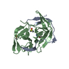
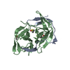
PDB-7h1p: PanDDA analysis group deposition -- Crystal Structure of ZIKV NS2B-NS3 protease in complex with Z425338146 Method: X-RAY DIFFRACTION / Resolution: 1.38 Å
PDB-7h1q: PanDDA analysis group deposition -- Crystal Structure of ZIKV NS2B-NS3 protease in complex with Z119990326 Method: X-RAY DIFFRACTION / Resolution: 1.411 Å
PDB-7h1r: PanDDA analysis group deposition -- Crystal Structure of ZIKV NS2B-NS3 protease in complex with Z270758780 Method: X-RAY DIFFRACTION / Resolution: 1.521 Å
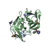
PDB-7h1s: PanDDA analysis group deposition -- Crystal Structure of ZIKV NS2B-NS3 protease in complex with Z57477251 Method: X-RAY DIFFRACTION / Resolution: 1.376 Å
PDB-7h1t: PanDDA analysis group deposition -- Crystal Structure of ZIKV NS2B-NS3 protease in complex with Z362020366 Method: X-RAY DIFFRACTION / Resolution: 1.419 Å
PDB-7h1u: PanDDA analysis group deposition -- Crystal Structure of ZIKV NS2B-NS3 protease in complex with Z212053854 Method: X-RAY DIFFRACTION / Resolution: 1.37 Å
PDB-7h1v: PanDDA analysis group deposition -- Crystal Structure of ZIKV NS2B-NS3 protease in complex with Z31113727 Method: X-RAY DIFFRACTION / Resolution: 1.36 Å
PDB-7h1w: PanDDA analysis group deposition -- Crystal Structure of ZIKV NS2B-NS3 protease in complex with Z270834034 Method: X-RAY DIFFRACTION / Resolution: 1.396 Å
PDB-7h1x: PanDDA analysis group deposition -- Crystal Structure of ZIKV NS2B-NS3 protease in complex with Z53833304 Method: X-RAY DIFFRACTION / Resolution: 1.398 Å
PDB-7h1y: PanDDA analysis group deposition -- Crystal Structure of ZIKV NS2B-NS3 protease in complex with Z57493554 Method: X-RAY DIFFRACTION / Resolution: 1.371 Å
PDB-7h1z: PanDDA analysis group deposition -- Crystal Structure of ZIKV NS2B-NS3 protease in complex with Z1198149728 Method: X-RAY DIFFRACTION / Resolution: 1.38 Å
PDB-7h20: PanDDA analysis group deposition -- Crystal Structure of ZIKV NS2B-NS3 protease in complex with Z4605084899 Method: X-RAY DIFFRACTION / Resolution: 1.39 Å
PDB-7h21: PanDDA analysis group deposition -- Crystal Structure of ZIKV NS2B-NS3 protease in complex with Z1269221363 Method: X-RAY DIFFRACTION / Resolution: 1.462 Å
PDB-7h22: PanDDA analysis group deposition -- Crystal Structure of ZIKV NS2B-NS3 protease in complex with Z1102357527 Method: X-RAY DIFFRACTION / Resolution: 1.554 Å
PDB-7h23: PanDDA analysis group deposition -- Crystal Structure of ZIKV NS2B-NS3 protease in complex with Z1148747945 Method: X-RAY DIFFRACTION / Resolution: 1.492 Å
PDB-7h24: PanDDA analysis group deposition -- Crystal Structure of ZIKV NS2B-NS3 protease in complex with Z1165350851 Method: X-RAY DIFFRACTION / Resolution: 1.64 Å
PDB-7h25: PanDDA analysis group deposition -- Crystal Structure of ZIKV NS2B-NS3 protease in complex with Z1198158918 Method: X-RAY DIFFRACTION / Resolution: 1.564 Å
PDB-7h26: PanDDA analysis group deposition -- Crystal Structure of ZIKV NS2B-NS3 protease in complex with Z1198317053 Method: X-RAY DIFFRACTION / Resolution: 1.849 Å
PDB-7h27: PanDDA analysis group deposition -- Crystal Structure of ZIKV NS2B-NS3 protease in complex with Z1203191681 Method: X-RAY DIFFRACTION / Resolution: 1.351 Å
PDB-7h28: PanDDA analysis group deposition -- Crystal Structure of ZIKV NS2B-NS3 protease in complex with Z1216833237 Method: X-RAY DIFFRACTION / Resolution: 1.799 Å
PDB-7h29: PanDDA analysis group deposition -- Crystal Structure of ZIKV NS2B-NS3 protease in complex with Z1269184613 Method: X-RAY DIFFRACTION / Resolution: 1.77 Å
PDB-7h2a: PanDDA analysis group deposition -- Crystal Structure of ZIKV NS2B-NS3 protease in complex with Z1269220427 Method: X-RAY DIFFRACTION / Resolution: 1.709 Å
PDB-7h2b: PanDDA analysis group deposition -- Crystal Structure of ZIKV NS2B-NS3 protease in complex with Z1272517105 Method: X-RAY DIFFRACTION / Resolution: 1.602 Å
PDB-7h2c: PanDDA analysis group deposition -- Crystal Structure of ZIKV NS2B-NS3 protease in complex with Z1328968520 Method: X-RAY DIFFRACTION / Resolution: 1.399 Å
PDB-7h2d: PanDDA analysis group deposition -- Crystal Structure of ZIKV NS2B-NS3 protease in complex with Z1397964787 Method: X-RAY DIFFRACTION / Resolution: 1.566 Å
PDB-7h2e: PanDDA analysis group deposition -- Crystal Structure of ZIKV NS2B-NS3 protease in complex with Z1428159350 Method: X-RAY DIFFRACTION / Resolution: 1.36 Å
PDB-7h2f: PanDDA analysis group deposition -- Crystal Structure of ZIKV NS2B-NS3 protease in complex with Z1491215378 Method: X-RAY DIFFRACTION / Resolution: 1.381 Å
PDB-7h2g: PanDDA analysis group deposition -- Crystal Structure of ZIKV NS2B-NS3 protease in complex with Z1509195674 Method: X-RAY DIFFRACTION / Resolution: 1.25 Å
PDB-7h2h: PanDDA analysis group deposition -- Crystal Structure of ZIKV NS2B-NS3 protease in complex with Z1575274523 Method: X-RAY DIFFRACTION / Resolution: 1.419 Å
PDB-7h2i: PanDDA analysis group deposition -- Crystal Structure of ZIKV NS2B-NS3 protease in complex with Z1587220559 Method: X-RAY DIFFRACTION / Resolution: 1.32 Å
PDB-7h2j: PanDDA analysis group deposition -- Crystal Structure of ZIKV NS2B-NS3 protease in complex with Z1787627869 Method: X-RAY DIFFRACTION / Resolution: 1.762 Å
PDB-7h2k: PanDDA analysis group deposition -- Crystal Structure of ZIKV NS2B-NS3 protease in complex with Z18769001 Method: X-RAY DIFFRACTION / Resolution: 1.595 Å
PDB-7h2l: PanDDA analysis group deposition -- Crystal Structure of ZIKV NS2B-NS3 protease in complex with Z19755216 Method: X-RAY DIFFRACTION / Resolution: 1.366 Å
PDB-7h2m: PanDDA analysis group deposition -- Crystal Structure of ZIKV NS2B-NS3 protease in complex with Z228586974 Method: X-RAY DIFFRACTION / Resolution: 1.712 Å
PDB-7h2n: PanDDA analysis group deposition -- Crystal Structure of ZIKV NS2B-NS3 protease in complex with Z228587394 Method: X-RAY DIFFRACTION / Resolution: 1.73 Å
PDB-7h2o: PanDDA analysis group deposition -- Crystal Structure of ZIKV NS2B-NS3 protease in complex with Z234898257 Method: X-RAY DIFFRACTION / Resolution: 1.707 Å
PDB-7h2p: PanDDA analysis group deposition -- Crystal Structure of ZIKV NS2B-NS3 protease in complex with Z2509342103 Method: X-RAY DIFFRACTION / Resolution: 1.452 Å
PDB-7h2q: PanDDA analysis group deposition -- Crystal Structure of ZIKV NS2B-NS3 protease in complex with Z270760338 Method: X-RAY DIFFRACTION / Resolution: 1.526 Å
PDB-7h2r: PanDDA analysis group deposition -- Crystal Structure of ZIKV NS2B-NS3 protease in complex with Z31735562 Method: X-RAY DIFFRACTION / Resolution: 1.321 Å
PDB-7h2s: PanDDA analysis group deposition -- Crystal Structure of ZIKV NS2B-NS3 protease in complex with Z396117078 Method: X-RAY DIFFRACTION / Resolution: 1.57 Å
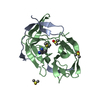
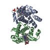
PDB-1aj7: IMMUNOGLOBULIN 48G7 GERMLINE FAB ANTIBODY COMPLEXED WITH HAPTEN 5-(PARA-NITROPHENYL PHOSPHONATE)-PENTANOIC ACID. AFFINITY MATURATION OF AN ESTEROLYTIC ANTIBODY
In the structure databanks used in Yorodumi, some data are registered as the other names, "COVID-19 virus" and "2019-nCoV". Here are the details of the virus and the list of structure data.
Jan 31, 2019. EMDB accession codes are about to change! (news from PDBe EMDB page)
EMDB accession codes are about to change! (news from PDBe EMDB page)
The allocation of 4 digits for EMDB accession codes will soon come to an end. Whilst these codes will remain in use, new EMDB accession codes will include an additional digit and will expand incrementally as the available range of codes is exhausted. The current 4-digit format prefixed with “EMD-” (i.e. EMD-XXXX) will advance to a 5-digit format (i.e. EMD-XXXXX), and so on. It is currently estimated that the 4-digit codes will be depleted around Spring 2019, at which point the 5-digit format will come into force.
The EM Navigator/Yorodumi systems omit the EMD- prefix.
Related info.:Q: What is EMD? / ID/Accession-code notation in Yorodumi/EM Navigator
 Movie
Movie Controller
 Structure viewers
 About Yorodumi Papers


 Authors
 External links
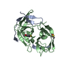


 Keywords
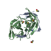
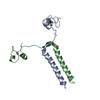
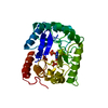
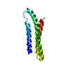
 zika virus
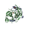
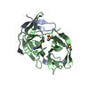