[English] 日本語
Yorodumi
- PDB-1aj4: STRUCTURE OF CALCIUM-SATURATED CARDIAC TROPONIN C, NMR, 1 STRUCTURE -

+
Open data


ID or keywords:

Loading...

-
Basic information

Entry
Database: PDB / ID: 1aj4
TitleSTRUCTURE OF CALCIUM-SATURATED CARDIAC TROPONIN C, NMR, 1 STRUCTURE
ComponentsTROPONIN C
KeywordsMUSCLE PROTEIN / CARDIAC / REGULATORY / CALCIUM BINDING
Function / homology
Function and homology information


cardiac Troponin complex / Striated Muscle Contraction / troponin I binding / skeletal muscle contraction / cardiac muscle contraction / calcium-dependent protein binding / calcium ion binding
Similarity search - Function
EF hand / EF-hand domain pair / EF-hand / Recoverin; domain 1 / EF-hand domain pair / EF-hand, calcium binding motif / EF-Hand 1, calcium-binding site / EF-hand calcium-binding domain. / EF-hand calcium-binding domain profile. / EF-hand domain ...EF hand / EF-hand domain pair / EF-hand / Recoverin; domain 1 / EF-hand domain pair / EF-hand, calcium binding motif / EF-Hand 1, calcium-binding site / EF-hand calcium-binding domain. / EF-hand calcium-binding domain profile. / EF-hand domain / EF-hand domain pair / Orthogonal Bundle / Mainly Alpha
Similarity search - Domain/homology
Troponin C, slow skeletal and cardiac muscles
Similarity search - Component
Biological speciesGallus gallus (chicken)
MethodSOLUTION NMR / simulated annealing
AuthorsSia, S.K. / Li, M.X. / Spyracopoulos, L. / Gagne, S.M. / Liu, W. / Putkey, J.A. / Sykes, B.D.
CitationJournal: J.Biol.Chem. / Year: 1997
Title: Structure of cardiac muscle troponin C unexpectedly reveals a closed regulatory domain.
Authors: Sia, S.K. / Li, M.X. / Spyracopoulos, L. / Gagne, S.M. / Liu, W. / Putkey, J.A. / Sykes, B.D.
History
DepositionMay 14, 1997Processing site: BNL
Revision 1.0May 20, 1998Provider: repository / Type: Initial release
Revision 1.1Mar 24, 2008Group: Version format compliance
Revision 1.2Jul 13, 2011Group: Version format compliance
Revision 1.3Nov 3, 2021Group: Database references / Derived calculations / Other
Category: database_2 / pdbx_database_status ...database_2 / pdbx_database_status / pdbx_struct_assembly / pdbx_struct_conn_angle / pdbx_struct_oper_list / struct_conn / struct_ref_seq_dif / struct_site
Item: _database_2.pdbx_DOI / _database_2.pdbx_database_accession ..._database_2.pdbx_DOI / _database_2.pdbx_database_accession / _pdbx_database_status.process_site / _pdbx_struct_conn_angle.ptnr1_auth_comp_id / _pdbx_struct_conn_angle.ptnr1_auth_seq_id / _pdbx_struct_conn_angle.ptnr1_label_atom_id / _pdbx_struct_conn_angle.ptnr1_label_comp_id / _pdbx_struct_conn_angle.ptnr1_label_seq_id / _pdbx_struct_conn_angle.ptnr3_auth_comp_id / _pdbx_struct_conn_angle.ptnr3_auth_seq_id / _pdbx_struct_conn_angle.ptnr3_label_atom_id / _pdbx_struct_conn_angle.ptnr3_label_comp_id / _pdbx_struct_conn_angle.ptnr3_label_seq_id / _pdbx_struct_conn_angle.value / _struct_conn.pdbx_dist_value / _struct_conn.ptnr1_auth_comp_id / _struct_conn.ptnr1_auth_seq_id / _struct_conn.ptnr1_label_asym_id / _struct_conn.ptnr1_label_atom_id / _struct_conn.ptnr1_label_comp_id / _struct_conn.ptnr1_label_seq_id / _struct_conn.ptnr2_auth_comp_id / _struct_conn.ptnr2_auth_seq_id / _struct_conn.ptnr2_label_asym_id / _struct_conn.ptnr2_label_atom_id / _struct_conn.ptnr2_label_comp_id / _struct_conn.ptnr2_label_seq_id / _struct_ref_seq_dif.details / _struct_site.pdbx_auth_asym_id / _struct_site.pdbx_auth_comp_id / _struct_site.pdbx_auth_seq_id

-
Structure visualization

Structure viewerMolecule:
MolmilJmol/JSmol

Downloads & links

-
Assembly

Deposited unit
A: TROPONIN C
hetero molecules


Theoretical massNumber of molelcules
Total (without water)18,4324
Polymers18,3111
Non-polymers1203
Water0
1


  • Idetical with deposited unit
  • defined by author
TypeNameSymmetry operationNumber
identity operation1_5551
NMR ensembles
DataCriteria
Number of conformers (submitted / calculated)1 / 35SMALLEST RMSD TO THE AVERAGE STRUCTURE
Representative

-
Components

#1: Protein TROPONIN C / / CTNC


Mass: 18311.275 Da / Num. of mol.: 1 / Mutation: C35S, C84S
Source method: isolated from a genetically manipulated source
Details: CARDIAC TROPONIN C, CALCIUM IONS BOUND AT SITES II, III AND IV
Source: (gene. exp.) Gallus gallus (chicken) / Tissue: MUSCLESkeletal muscle / Cell line: BL21 / Cellular location: THIN FILAMENTMyofilament / Gene: CTNC(A-CYS) / Organ: HEART / Plasmid: PET-23D / Gene (production host): CTNC(A-CYS) / Production host: Escherichia coli (E. coli) / Strain (production host): BL21 (DE3) PLYSS / References: UniProt: P09860
#2: Chemical ChemComp-CA / CALCIUM ION


Mass: 40.078 Da / Num. of mol.: 3 / Source method: obtained synthetically / Formula: Ca

-
Experimental details

-
Experiment

ExperimentMethod: SOLUTION NMR
NMR experiment
Conditions-IDExperiment-IDSolution-IDType
111NOESY
121TOCSY

-
Sample preparation

Sample conditionspH: 6.7 / Temperature: 303 K
Crystal grow
*PLUS
Method: other / Details: NMR

-
NMR measurement

NMR spectrometer
TypeManufacturerModelField strength (MHz)Spectrometer-ID
Varian UNITYPLUSVarianUNITYPLUS5001
Varian UNITYPLUSVarianUNITYPLUS6002

-
Processing

Software
NameVersionClassification
X-PLOR3.1model building
X-PLOR3.1refinement
X-PLOR3.1phasing
NMR software
NameVersionDeveloperClassification
X-PLOR3.1BRUNGERrefinement
X-PLORstructure solution
Refinement
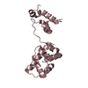
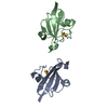
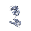
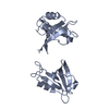
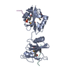
Method: simulated annealing / Software ordinal: 1
Details: CALCULATIONS OF THE N- AND C-DOMAINS WERE PERFORMED SEPARATELY BECAUSE THE CENTRAL LINKER IS UNSTRUCTURED AND FLEXIBLE IN SOLUTION. THE ORIENTATION OF THE TWO DOMAINS WITH RESPECT TO EACH ...Details: CALCULATIONS OF THE N- AND C-DOMAINS WERE PERFORMED SEPARATELY BECAUSE THE CENTRAL LINKER IS UNSTRUCTURED AND FLEXIBLE IN SOLUTION. THE ORIENTATION OF THE TWO DOMAINS WITH RESPECT TO EACH OTHER AND THE STRUCTURE OF THE CENTRAL LINKER (RESIDUES 86 - 94) ARE THEREFORE UNDEFINED. THE N-DOMAIN (2 - 85) AND C-DOMAIN (95 - 161) IN THIS STRUCTURE ARE THE STRUCTURES IN THE ENSEMBLES CLOSEST TO THE UNMINIMIZED AVERAGE STRUCTURES. IN PARTICULAR, THE N-DOMAIN HERE CORRESPONDS TO MODEL 25 IN 2CTN, AND THE C-DOMAIN TO MODEL 3 IN 3CTN. THE CENTRAL LINKER HERE IS THE RESULT OF ENERGY MINIMIZATION WITHOUT EXPERIMENTAL RESTRAINTS, AND IS ADDED ONLY AS A TOOL OF CONVENIENCE.
NMR ensembleConformer selection criteria: SMALLEST RMSD TO THE AVERAGE STRUCTURE
Conformers calculated total number: 35 / Conformers submitted total number: 1

+
About Yorodumi

-
News

-
Feb 9, 2022. New format data for meta-information of EMDB entries

New format data for meta-information of EMDB entries

  • Version 3 of the EMDB header file is now the official format.
  • The previous official version 1.9 will be removed from the archive.

Related info.:EMDB header

External links:wwPDB to switch to version 3 of the EMDB data model

-
Aug 12, 2020. Covid-19 info

Covid-19 info

URL: https://pdbj.org/emnavi/covid19.php

New page: Covid-19 featured information page in EM Navigator.

Related info.:Covid-19 info / Mar 5, 2020. Novel coronavirus structure data

+
Mar 5, 2020. Novel coronavirus structure data

Novel coronavirus structure data

Related info.:Yorodumi Speices / Aug 12, 2020. Covid-19 info

External links:COVID-19 featured content - PDBj / Molecule of the Month (242):Coronavirus Proteases

+
Jan 31, 2019. EMDB accession codes are about to change! (news from PDBe EMDB page)

EMDB accession codes are about to change! (news from PDBe EMDB page)

  • The allocation of 4 digits for EMDB accession codes will soon come to an end. Whilst these codes will remain in use, new EMDB accession codes will include an additional digit and will expand incrementally as the available range of codes is exhausted. The current 4-digit format prefixed with “EMD-” (i.e. EMD-XXXX) will advance to a 5-digit format (i.e. EMD-XXXXX), and so on. It is currently estimated that the 4-digit codes will be depleted around Spring 2019, at which point the 5-digit format will come into force.
  • The EM Navigator/Yorodumi systems omit the EMD- prefix.

Related info.:Q: What is EMD? / ID/Accession-code notation in Yorodumi/EM Navigator

External links:EMDB Accession Codes are Changing Soon! / Contact to PDBj

+
Jul 12, 2017. Major update of PDB

Major update of PDB

  • wwPDB released updated PDB data conforming to the new PDBx/mmCIF dictionary.
  • This is a major update changing the version number from 4 to 5, and with Remediation, in which all the entries are updated.
  • In this update, many items about electron microscopy experimental information are reorganized (e.g. em_software).
  • Now, EM Navigator and Yorodumi are based on the updated data.

External links:wwPDB Remediation / Enriched Model Files Conforming to OneDep Data Standards Now Available in the PDB FTP Archive

-
Yorodumi

Thousand views of thousand structures

  • Yorodumi is a browser for structure data from EMDB, PDB, SASBDB, etc.
  • This page is also the successor to EM Navigator detail page, and also detail information page/front-end page for Omokage search.
  • The word "yorodu" (or yorozu) is an old Japanese word meaning "ten thousand". "mi" (miru) is to see.

Related info.:EMDB / PDB / SASBDB / Comparison of 3 databanks / Yorodumi Search / Aug 31, 2016. New EM Navigator & Yorodumi / Yorodumi Papers / Jmol/JSmol / Function and homology information / Changes in new EM Navigator and Yorodumi

Read more