[English] 日本語
Yorodumi Papers
- Database of articles cited by EMDB/PDB/SASBDB data -

+
Search query

Keywords
Structure methods
Author
Journal
IF

-
Structure paper

TitleThe myosin X motor is optimized for movement on actin bundles.
Journal, issue, pagesNat Commun, Vol. 7, Page 12456, Year 2016
Publish dateSep 1, 2016
AuthorsVirginie Ropars / Zhaohui Yang / Tatiana Isabet / Florian Blanc / Kaifeng Zhou / Tianming Lin / Xiaoyan Liu / Pascale Hissier / Frédéric Samazan / Béatrice Amigues / Eric D Yang / Hyokeun Park / Olena Pylypenko / Marco Cecchini / Charles V Sindelar / H Lee Sweeney / Anne Houdusse /
PubMed AbstractMyosin X has features not found in other myosins. Its structure must underlie its unique ability to generate filopodia, which are essential for neuritogenesis, wound healing, cancer metastasis and ...Myosin X has features not found in other myosins. Its structure must underlie its unique ability to generate filopodia, which are essential for neuritogenesis, wound healing, cancer metastasis and some pathogenic infections. By determining high-resolution structures of key components of this motor, and characterizing the in vitro behaviour of the native dimer, we identify the features that explain the myosin X dimer behaviour. Single-molecule studies demonstrate that a native myosin X dimer moves on actin bundles with higher velocities and takes larger steps than on single actin filaments. The largest steps on actin bundles are larger than previously reported for artificially dimerized myosin X constructs or any other myosin. Our model and kinetic data explain why these large steps and high velocities can only occur on bundled filaments. Thus, myosin X functions as an antiparallel dimer in cells with a unique geometry optimized for movement on actin bundles.
External linksNat Commun / PubMed:27580874 / PubMed Central
MethodsEM (helical sym.) / X-ray diffraction
Resolution1.8 - 9.1 Å
Structure data

EMDB-8244, PDB-5kg8:
Rigor myosin X co-complexed with an actin filament
Method: EM (helical sym.) / Resolution: 9.1 Å

PDB-5hmo:
myosin X motor activity
Method: X-RAY DIFFRACTION / Resolution: 3.493 Å

PDB-5hmp:
Myosin Vc pre-powerstroke state
Method: X-RAY DIFFRACTION / Resolution: 2.397 Å

PDB-5i0h:
Crystal structure of myosin X motor domain in pre-powerstroke state
Method: X-RAY DIFFRACTION / Resolution: 1.8 Å

PDB-5i0i:
Crystal structure of myosin X motor domain with 2IQ motifs in pre-powerstroke state
Method: X-RAY DIFFRACTION / Resolution: 3.15 Å

Chemicals

ChemComp-MG:
Unknown entry

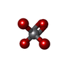
ChemComp-VO4:
VANADATE ION / Vanadate

ChemComp-ADP:
ADENOSINE-5'-DIPHOSPHATE / ADP, energy-carrying molecule*YM / Adenosine diphosphate

ChemComp-EDO:
1,2-ETHANEDIOL / Ethylene glycol

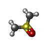
ChemComp-DMS:
DIMETHYL SULFOXIDE / DMSO, precipitant*YM / Dimethyl sulfoxide

ChemComp-HOH:
WATER / Water

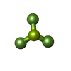
ChemComp-BEF:
BERYLLIUM TRIFLUORIDE ION

ChemComp-SO4:
SULFATE ION / Sulfate

ChemComp-MPO:
3[N-MORPHOLINO]PROPANE SULFONIC ACID / pH buffer*YM / MOPS

Source
  • homo sapiens (human)
  • oryctolagus cuniculus (rabbit)
  • bos taurus (cattle)
KeywordsMOTOR PROTEIN / myosin / motor domain / molecular motor / pre-powerstroke state / motility / pre-powerstrocke state / myosin molecular motors cytoskeletal motility

+
About Yorodumi Papers

-
News

-
Feb 9, 2022. New format data for meta-information of EMDB entries

New format data for meta-information of EMDB entries

  • Version 3 of the EMDB header file is now the official format.
  • The previous official version 1.9 will be removed from the archive.

Related info.:EMDB header

External links:wwPDB to switch to version 3 of the EMDB data model

-
Aug 12, 2020. Covid-19 info

Covid-19 info

URL: https://pdbj.org/emnavi/covid19.php

New page: Covid-19 featured information page in EM Navigator.

Related info.:Covid-19 info / Mar 5, 2020. Novel coronavirus structure data

+
Mar 5, 2020. Novel coronavirus structure data

Novel coronavirus structure data

Related info.:Yorodumi Speices / Aug 12, 2020. Covid-19 info

External links:COVID-19 featured content - PDBj / Molecule of the Month (242):Coronavirus Proteases

+
Jan 31, 2019. EMDB accession codes are about to change! (news from PDBe EMDB page)

EMDB accession codes are about to change! (news from PDBe EMDB page)

  • The allocation of 4 digits for EMDB accession codes will soon come to an end. Whilst these codes will remain in use, new EMDB accession codes will include an additional digit and will expand incrementally as the available range of codes is exhausted. The current 4-digit format prefixed with “EMD-” (i.e. EMD-XXXX) will advance to a 5-digit format (i.e. EMD-XXXXX), and so on. It is currently estimated that the 4-digit codes will be depleted around Spring 2019, at which point the 5-digit format will come into force.
  • The EM Navigator/Yorodumi systems omit the EMD- prefix.

Related info.:Q: What is EMD? / ID/Accession-code notation in Yorodumi/EM Navigator

External links:EMDB Accession Codes are Changing Soon! / Contact to PDBj

+
Jul 12, 2017. Major update of PDB

Major update of PDB

  • wwPDB released updated PDB data conforming to the new PDBx/mmCIF dictionary.
  • This is a major update changing the version number from 4 to 5, and with Remediation, in which all the entries are updated.
  • In this update, many items about electron microscopy experimental information are reorganized (e.g. em_software).
  • Now, EM Navigator and Yorodumi are based on the updated data.

External links:wwPDB Remediation / Enriched Model Files Conforming to OneDep Data Standards Now Available in the PDB FTP Archive

-
Yorodumi Papers

Database of articles cited by EMDB/PDB/SASBDB data

  • Database of articles cited by EMDB, PDB, and SASBDB entries
  • Using PubMed data

Related info.:EMDB / PDB / SASBDB / Yorodumi / EMN Papers / Changes in new EM Navigator and Yorodumi

Read more