 Movie
Movie Controller
 Structure viewers
 About Yorodumi Papers


+Search query
-Structure paper
| Title | Structural insights into lipid membrane binding by human ferlins. |
|---|---|
| Journal, issue, pages | EMBO J, Vol. 44, Issue 14, Page 3926-3958, Year 2025 |
| Publish date | May 28, 2025 |
 Authors Authors | Constantin Cretu / Aleksandar Chernev / Csaba Zoltán Kibédi Szabó / Vladimir Pena / Henning Urlaub / Tobias Moser / Julia Preobraschenski /   |
| PubMed Abstract | Ferlins are ancient membrane proteins with a unique architecture, and play central roles in crucial processes that involve Ca-dependent vesicle fusion. Despite their links to multiple human diseases ...Ferlins are ancient membrane proteins with a unique architecture, and play central roles in crucial processes that involve Ca-dependent vesicle fusion. Despite their links to multiple human diseases and numerous functional studies, a mechanistic understanding of how these multi-C domain-containing proteins interact with lipid membranes to promote membrane remodelling and fusion is currently lacking. Here we obtain near-complete cryo-electron microscopy structures of human myoferlin and dysferlin in their Ca- and lipid-bound states. We show that ferlins adopt compact, ring-like tertiary structures upon membrane binding. The top arch of the ferlin ring, composed of the CC-CD region, is rigid and exhibits only little variability across the observed functional states. In contrast, the N-terminal CB and the C-terminal CF-CG domains cycle between alternative conformations and, in response to Ca, close the ferlin ring, promoting tight interaction with the target membrane. Probing key domain interfaces validates the observed architecture, and informs a model of how ferlins engage lipid bilayers in a Ca-dependent manner. This work reveals the general principles of human ferlin structures and provides a framework for future analyses of ferlin-dependent cellular functions and disease mechanisms. |
 External links External links | EMBO J / PubMed:40437073 / PubMed Central |
| Methods | EM (single particle) |
| Resolution | 2.56 - 3.54 Å |
| Structure data | EMDB-51902, PDB-9h6x: EMDB-53222, PDB-9qkv: EMDB-53225, PDB-9qle: EMDB-53226, PDB-9qlf: EMDB-53229, PDB-9qln: EMDB-53233, PDB-9qls: |
| Chemicals | 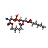 ChemComp-PSF:  ChemComp-CA: |
| Source |
|
 Keywords Keywords | MEMBRANE PROTEIN / Ferlin / multi-C2 domain protein / tail-anchored membrane protein / vesicle docking and fusion / membrane remodeling / Ferlins / myoferlin / multi-C2 domains / lipid nanodisc / multi-C2 domain / dysferlin |


 homo sapiens (human)