 Movie
Movie Controller

[English] 日本語
 Yorodumi
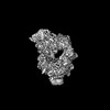
- PDB-9qkv: Human myoferlin (1-1997) in complex with an MSP2N2 lipid nanodisc... -
+ Open data

- Basic information
| Entry | Database: PDB / ID: 9qkv | ||||||
|---|---|---|---|---|---|---|---|
| Title | Human myoferlin (1-1997) in complex with an MSP2N2 lipid nanodisc (15 mol% DOPS, 5 mol% Cholesterol) | ||||||
 Components Components | Myoferlin | ||||||
 Keywords Keywords | MEMBRANE PROTEIN / Ferlins / myoferlin / multi-C2 domains / lipid nanodisc | ||||||
| Function / homology |  Function and homology information Function and homology informationregulation of neurotransmitter secretion / plasma membrane repair / blood circulation / centriolar satellite / ciliary tip / muscle contraction / phospholipid binding / caveola / nuclear envelope / late endosome membrane ...regulation of neurotransmitter secretion / plasma membrane repair / blood circulation / centriolar satellite / ciliary tip / muscle contraction / phospholipid binding / caveola / nuclear envelope / late endosome membrane / synaptic vesicle membrane / nuclear membrane / cytoplasmic vesicle / cilium / ciliary basal body / calcium ion binding / extracellular exosome / plasma membrane Similarity search - Function | ||||||
| Biological species |  Homo sapiens (human) Homo sapiens (human) | ||||||
| Method | ELECTRON MICROSCOPY / single particle reconstruction / cryo EM / Resolution: 2.74 Å | ||||||
 Authors Authors | Cretu, C. / Moser, T. | ||||||
| Funding support |  Germany, 1items Germany, 1items
| ||||||
 Citation Citation | Journal: EMBO J / Year: 2025 Title: Structural insights into lipid membrane binding by human ferlins. Authors: Constantin Cretu / Aleksandar Chernev / Csaba Zoltán Kibédi Szabó / Vladimir Pena / Henning Urlaub / Tobias Moser / Julia Preobraschenski /  Abstract: Ferlins are ancient membrane proteins with a unique architecture, and play central roles in crucial processes that involve Ca-dependent vesicle fusion. Despite their links to multiple human diseases ...Ferlins are ancient membrane proteins with a unique architecture, and play central roles in crucial processes that involve Ca-dependent vesicle fusion. Despite their links to multiple human diseases and numerous functional studies, a mechanistic understanding of how these multi-C domain-containing proteins interact with lipid membranes to promote membrane remodelling and fusion is currently lacking. Here we obtain near-complete cryo-electron microscopy structures of human myoferlin and dysferlin in their Ca- and lipid-bound states. We show that ferlins adopt compact, ring-like tertiary structures upon membrane binding. The top arch of the ferlin ring, composed of the CC-CD region, is rigid and exhibits only little variability across the observed functional states. In contrast, the N-terminal CB and the C-terminal CF-CG domains cycle between alternative conformations and, in response to Ca, close the ferlin ring, promoting tight interaction with the target membrane. Probing key domain interfaces validates the observed architecture, and informs a model of how ferlins engage lipid bilayers in a Ca-dependent manner. This work reveals the general principles of human ferlin structures and provides a framework for future analyses of ferlin-dependent cellular functions and disease mechanisms. | ||||||
| History |
|
- Structure visualization
| Structure viewer | Molecule: MolmilJmol/JSmol |
|---|
- Downloads & links
-Download
| PDBx/mmCIF format | 9qkv.cif.gz | 451.2 KB | Display | PDBx/mmCIF format |
|---|---|---|---|---|
| PDB format | pdb9qkv.ent.gz | Display | PDB format | |
| PDBx/mmJSON format | 9qkv.json.gz | Tree view | PDBx/mmJSON format | |
| Others |  Other downloads Other downloads |
-Validation report
| Arichive directory | https://data.pdbj.org/pub/pdb/validation_reports/qk/9qkvftp://data.pdbj.org/pub/pdb/validation_reports/qk/9qkv | HTTPS FTP |
|---|
-Related structure data
| Related structure data |  53222MC  9h6xC  9qleC  9qlfC  9qlnC  9qlsC C: citing same article ( M: map data used to model this data |
|---|---|
| Similar structure data |


-Links
 PDBj
PDBj


- Assembly
| Deposited unit | 
|
|---|---|
| 1 |
|
-Components
| #1: Protein | Mass: 232625.953 Da / Num. of mol.: 1 Source method: isolated from a genetically manipulated source Details: The cytosolic domain of human myoferlin (residues 1-1997), cloned in-frame with a twin-StrepII affinity tag. Source: (gene. exp.) Homo sapiens (human) / Gene: MYOF, FER1L3, KIAA1207 / Plasmid: pCC76Details (production host): twin-StrepII-HRV3C-Myoferlin(1-1997) Cell (production host): Sf9 cells / Production host:   Spodoptera frugiperda (fall armyworm) / References: UniProt: Q9NZM1 Spodoptera frugiperda (fall armyworm) / References: UniProt: Q9NZM1 | ||||||
|---|---|---|---|---|---|---|---|
| #2: Chemical | 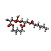  Mass: 455.437 Da / Num. of mol.: 2 / Source method: obtained synthetically / Formula: C18H34NO10P / Feature type: SUBJECT OF INVESTIGATION Mass: 455.437 Da / Num. of mol.: 2 / Source method: obtained synthetically / Formula: C18H34NO10P / Feature type: SUBJECT OF INVESTIGATION#3: Chemical | ChemComp-CA /   Mass: 40.078 Da / Num. of mol.: 10 / Source method: obtained synthetically / Formula: Ca / Feature type: SUBJECT OF INVESTIGATION Mass: 40.078 Da / Num. of mol.: 10 / Source method: obtained synthetically / Formula: Ca / Feature type: SUBJECT OF INVESTIGATIONHas ligand of interest | Y | Has protein modification | N | |
-Experimental details
-Experiment
| Experiment | Method: ELECTRON MICROSCOPY |
|---|---|
| EM experiment | Aggregation state: PARTICLE / 3D reconstruction method: single particle reconstruction |
- Sample preparation
| Component | Name: Human myoferlin (1-1997) in complex with an MSP2N2 lipid nanodisc (comprising 15 mol% DOPS, 5 mol% Cholesterol) Type: COMPLEX / Entity ID: #1 / Source: RECOMBINANT | ||||||||||||||||||||||||||||||
|---|---|---|---|---|---|---|---|---|---|---|---|---|---|---|---|---|---|---|---|---|---|---|---|---|---|---|---|---|---|---|---|
| Molecular weight | Experimental value: NO | ||||||||||||||||||||||||||||||
| Source (natural) | Organism: Homo sapiens (human) | ||||||||||||||||||||||||||||||
| Source (recombinant) | Organism: Spodoptera frugiperda (fall armyworm) | ||||||||||||||||||||||||||||||
| Buffer solution | pH: 7.5 | ||||||||||||||||||||||||||||||
| Buffer component |
| ||||||||||||||||||||||||||||||
| Specimen | Conc.: 0.77 mg/ml / Embedding applied: NO / Shadowing applied: NO / Staining applied: NO / Vitrification applied: YES Details: Prior to cryo-EM grid preparation, the sample was crosslinked with glutaraldehyde (0.05 % (v/v)) in batch and purified by size-exclusion chromatography | ||||||||||||||||||||||||||||||
| Specimen support | Details: The grid was treated with the Plasma Cleaner (Harrick Plasma) for 1 min at medium settings, followed by vitrification in liquid ethane-propane (37%/63%), cooled by liquid nitrogen Grid material: COPPER / Grid mesh size: 200 divisions/in. / Grid type: Quantifoil R1.2/1.3 | ||||||||||||||||||||||||||||||
| Vitrification | Instrument: FEI VITROBOT MARK IV / Cryogen name: ETHANE-PROPANE / Humidity: 100 % / Chamber temperature: 277.15 K |
- Electron microscopy imaging
| Experimental equipment |  Model: Titan Krios / Image courtesy: FEI Company |
|---|---|
| Microscopy | Model: TFS KRIOS |
| Electron gun | Electron source:  FIELD EMISSION GUN / Accelerating voltage: 300 kV / Illumination mode: FLOOD BEAM FIELD EMISSION GUN / Accelerating voltage: 300 kV / Illumination mode: FLOOD BEAM |
| Electron lens | Mode: BRIGHT FIELD / Nominal magnification: 165000 X / Nominal defocus max: 2000 nm / Nominal defocus min: 800 nm / Cs: 2.7 mm / C2 aperture diameter: 50 µm / Alignment procedure: COMA FREE |
| Specimen holder | Cryogen: NITROGEN / Specimen holder model: FEI TITAN KRIOS AUTOGRID HOLDER |
| Image recording | Average exposure time: 3.45 sec. / Electron dose: 39.85 e/Å2 / Film or detector model: FEI FALCON IV (4k x 4k) / Num. of grids imaged: 1 / Num. of real images: 9800 / Details: 9621 movies were accepted after curation |
| EM imaging optics | Energyfilter name: TFS Selectris / Energyfilter slit width: 10 eV |
- Processing
| EM software |
| ||||||||||||||||||||||||||||||||||||
|---|---|---|---|---|---|---|---|---|---|---|---|---|---|---|---|---|---|---|---|---|---|---|---|---|---|---|---|---|---|---|---|---|---|---|---|---|---|
| CTF correction | Type: PHASE FLIPPING AND AMPLITUDE CORRECTION | ||||||||||||||||||||||||||||||||||||
| Particle selection | Num. of particles selected: 2917286 | ||||||||||||||||||||||||||||||||||||
| Symmetry | Point symmetry: C1 (asymmetric) | ||||||||||||||||||||||||||||||||||||
| 3D reconstruction | Resolution: 2.74 Å / Resolution method: FSC 0.143 CUT-OFF / Num. of particles: 277729 / Num. of class averages: 1 / Symmetry type: POINT | ||||||||||||||||||||||||||||||||||||
| Atomic model building | B value: 135.43 / Protocol: AB INITIO MODEL / Space: REAL | ||||||||||||||||||||||||||||||||||||
| Atomic model building | Source name: AlphaFold / Type: in silico model | ||||||||||||||||||||||||||||||||||||
| Refinement | Cross valid method: NONE Stereochemistry target values: GeoStd + Monomer Library + CDL v1.2 | ||||||||||||||||||||||||||||||||||||
| Displacement parameters | Biso mean: 122.5 Å2 | ||||||||||||||||||||||||||||||||||||
| Refine LS restraints |
|
