 Movie
Movie Controller
 Structure viewers
 About Yorodumi Papers


+Search query
-Structure paper
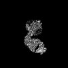
| Title | Fusion protein strategies for cryo-EM study of G protein-coupled receptors. |
|---|---|
| Journal, issue, pages | Nat Commun, Vol. 13, Issue 1, Page 4366, Year 2022 |
| Publish date | Jul 28, 2022 |
 Authors Authors | Kaihua Zhang / Hao Wu / Nicholas Hoppe / Aashish Manglik / Yifan Cheng /  |
| PubMed Abstract | Single particle cryogenic-electron microscopy (cryo-EM) is used extensively to determine structures of activated G protein-coupled receptors (GPCRs) in complex with G proteins or arrestins. However, ...Single particle cryogenic-electron microscopy (cryo-EM) is used extensively to determine structures of activated G protein-coupled receptors (GPCRs) in complex with G proteins or arrestins. However, applying it to GPCRs without signaling proteins remains challenging because most receptors lack structural features in their soluble domains to facilitate image alignment. In GPCR crystallography, inserting a fusion protein between transmembrane helices 5 and 6 is a highly successful strategy for crystallization. Although a similar strategy has the potential to broadly facilitate cryo-EM structure determination of GPCRs alone without signaling protein, the critical determinants that make this approach successful are not yet clear. Here, we address this shortcoming by exploring different fusion protein designs, which lead to structures of antagonist bound A adenosine receptor at 3.4 Å resolution and unliganded Smoothened at 3.7 Å resolution. The fusion strategies explored here are likely applicable to cryo-EM interrogation of other GPCRs and small integral membrane proteins. |
 External links External links | Nat Commun / PubMed:35902590 / PubMed Central |
| Methods | EM (single particle) |
| Resolution | 3.4 - 6.7 Å |
| Structure data | EMDB-25648, PDB-7t32: EMDB-27062, PDB-8cxo:  EMDB-27063: Cryo-EM structure of the unliganded mSMO-PGS1 in a lipidic environment |
| Chemicals | 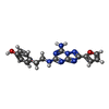 ChemComp-ZMA:  ChemComp-CLR: |
| Source |
|
 Keywords Keywords | MEMBRANE PROTEIN / A2AAR / GPCR / ADENOSINE RECEPTOR / G-protein coupled receptor / Smoothened / unliganded state / lipid system |


 homo sapiens (human)

 pyrococcus abyssi (strain ge5 / orsay) (archaea)
