 Movie
Movie Controller

[English] 日本語
 Yorodumi
- EMDB-25648: CryoEM structure of the adenosine 2A receptor-BRIL/Anti BRIL Fab ... -
+ Open data

- Basic information
| Entry |  | |||||||||
|---|---|---|---|---|---|---|---|---|---|---|
| Title | CryoEM structure of the adenosine 2A receptor-BRIL/Anti BRIL Fab complex with ZM241385 | |||||||||
 Map data Map data | Structure of the adenosine 2A receptor-BRIL/Anti BRIL Fab with ZM241385 | |||||||||
 Sample Sample |
| |||||||||
 Keywords Keywords | A2AAR / GPCR / ADENOSINE RECEPTOR / MEMBRANE PROTEIN | |||||||||
| Function / homology |  Function and homology information Function and homology informationregulation of norepinephrine secretion / negative regulation of alpha-beta T cell activation / positive regulation of circadian sleep/wake cycle, sleep / Adenosine P1 receptors / positive regulation of acetylcholine secretion, neurotransmission / G protein-coupled adenosine receptor activity / response to purine-containing compound / G protein-coupled adenosine receptor signaling pathway / NGF-independant TRKA activation / Surfactant metabolism ...regulation of norepinephrine secretion / negative regulation of alpha-beta T cell activation / positive regulation of circadian sleep/wake cycle, sleep / Adenosine P1 receptors / positive regulation of acetylcholine secretion, neurotransmission / G protein-coupled adenosine receptor activity / response to purine-containing compound / G protein-coupled adenosine receptor signaling pathway / NGF-independant TRKA activation / Surfactant metabolism / synaptic transmission, dopaminergic / type 5 metabotropic glutamate receptor binding / negative regulation of vascular permeability / synaptic transmission, cholinergic / intermediate filament / presynaptic active zone / positive regulation of urine volume / response to caffeine / blood circulation / sensory perception / positive regulation of glutamate secretion / eating behavior / inhibitory postsynaptic potential / regulation of calcium ion transport / alpha-actinin binding / asymmetric synapse / axolemma / membrane depolarization / cellular defense response / prepulse inhibition / phagocytosis / neuron projection morphogenesis / positive regulation of synaptic transmission, glutamatergic / astrocyte activation / presynaptic modulation of chemical synaptic transmission / positive regulation of long-term synaptic potentiation / positive regulation of synaptic transmission, GABAergic / positive regulation of protein secretion / central nervous system development / response to amphetamine / regulation of mitochondrial membrane potential / positive regulation of apoptotic signaling pathway / apoptotic signaling pathway / synaptic transmission, glutamatergic / excitatory postsynaptic potential / locomotory behavior / electron transport chain / negative regulation of inflammatory response / vasodilation / adenylate cyclase-modulating G protein-coupled receptor signaling pathway / blood coagulation / cell-cell signaling / adenylate cyclase-activating G protein-coupled receptor signaling pathway / presynaptic membrane / G alpha (s) signalling events / phospholipase C-activating G protein-coupled receptor signaling pathway / negative regulation of neuron apoptotic process / calmodulin binding / positive regulation of ERK1 and ERK2 cascade / periplasmic space / electron transfer activity / postsynaptic membrane / iron ion binding / response to xenobiotic stimulus / inflammatory response / negative regulation of cell population proliferation / neuronal cell body / heme binding / apoptotic process / regulation of DNA-templated transcription / lipid binding / dendrite / protein-containing complex binding / glutamatergic synapse / enzyme binding / membrane / identical protein binding / plasma membrane Similarity search - Function | |||||||||
| Biological species |  Homo sapiens (human) Homo sapiens (human) | |||||||||
| Method | single particle reconstruction / cryo EM / Resolution: 3.4 Å | |||||||||
 Authors Authors | Zhang KH / Wu H / Hoppe N / Manglik A / Cheng YF | |||||||||
| Funding support |  United States, 1 items United States, 1 items
| |||||||||
 Citation Citation | Journal: Nat Commun / Year: 2022 Title: Fusion protein strategies for cryo-EM study of G protein-coupled receptors. Authors: Kaihua Zhang / Hao Wu / Nicholas Hoppe / Aashish Manglik / Yifan Cheng / Abstract: Single particle cryogenic-electron microscopy (cryo-EM) is used extensively to determine structures of activated G protein-coupled receptors (GPCRs) in complex with G proteins or arrestins. However, ...Single particle cryogenic-electron microscopy (cryo-EM) is used extensively to determine structures of activated G protein-coupled receptors (GPCRs) in complex with G proteins or arrestins. However, applying it to GPCRs without signaling proteins remains challenging because most receptors lack structural features in their soluble domains to facilitate image alignment. In GPCR crystallography, inserting a fusion protein between transmembrane helices 5 and 6 is a highly successful strategy for crystallization. Although a similar strategy has the potential to broadly facilitate cryo-EM structure determination of GPCRs alone without signaling protein, the critical determinants that make this approach successful are not yet clear. Here, we address this shortcoming by exploring different fusion protein designs, which lead to structures of antagonist bound A adenosine receptor at 3.4 Å resolution and unliganded Smoothened at 3.7 Å resolution. The fusion strategies explored here are likely applicable to cryo-EM interrogation of other GPCRs and small integral membrane proteins. #1: Journal: Science / Year: 2012Title: Structural Basis for Allosteric Regulation of GPCRs by Sodium Ions Authors: Liu W | |||||||||
| History |
|
- Structure visualization
| Supplemental images |
|---|

- Downloads & links
-EMDB archive
| Map data | emd_25648.map.gz | 97.2 MB | EMDB map data format | |
|---|---|---|---|---|
| Header (meta data) | emd-25648-v30.xmlemd-25648.xml | 10.8 KB 10.8 KB | Display Display | EMDB header |
| Images | 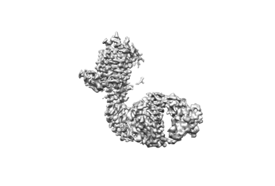 emd_25648.png emd_25648.png | 33.5 KB | ||
| Filedesc metadata | emd-25648.cif.gz | 5.4 KB | ||
| Archive directory |  http://ftp.pdbj.org/pub/emdb/structures/EMD-25648ftp://ftp.pdbj.org/pub/emdb/structures/EMD-25648 http://ftp.pdbj.org/pub/emdb/structures/EMD-25648ftp://ftp.pdbj.org/pub/emdb/structures/EMD-25648 | HTTPS FTP |
-Related structure data
| Related structure data |  7t32MC  8cxoC M: atomic model generated by this map C: citing same article ( |
|---|---|
| Similar structure data |


-Links
| EMDB pages | EMDB (EBI/PDBe) / EMDataResource |
|---|---|
| Related items in Molecule of the Month |


-Map
| File | Download / File: emd_25648.map.gz / Format: CCP4 / Size: 103 MB / Type: IMAGE STORED AS FLOATING POINT NUMBER (4 BYTES) | ||||||||||||||||||||||||||||||||||||
|---|---|---|---|---|---|---|---|---|---|---|---|---|---|---|---|---|---|---|---|---|---|---|---|---|---|---|---|---|---|---|---|---|---|---|---|---|---|
| Annotation | Structure of the adenosine 2A receptor-BRIL/Anti BRIL Fab with ZM241385 | ||||||||||||||||||||||||||||||||||||
| Projections & slices | Image control
Images are generated by Spider. | ||||||||||||||||||||||||||||||||||||
| Voxel size | X=Y=Z: 0.835 Å | ||||||||||||||||||||||||||||||||||||
| Density |
| ||||||||||||||||||||||||||||||||||||
| Symmetry | Space group: 1 | ||||||||||||||||||||||||||||||||||||
| Details | EMDB XML:
|
 Z (Sec.)
 Y (Row.)
 X (Col.)


-Supplemental data
- Sample components
-Entire : A2A adenosine receptor-BRIL/Anti BRIL Fab complex
| Entire | Name: A2A adenosine receptor-BRIL/Anti BRIL Fab complex |
|---|---|
| Components |
|
-Supramolecule #1: A2A adenosine receptor-BRIL/Anti BRIL Fab complex
| Supramolecule | Name: A2A adenosine receptor-BRIL/Anti BRIL Fab complex / type: complex / ID: 1 / Parent: 0 / Macromolecule list: #1 |
|---|---|
| Source (natural) | Organism: Homo sapiens (human) |
-Macromolecule #1: Adenosine receptor A2a/Soluble cytochrome b562 Fusion Protein
| Macromolecule | Name: Adenosine receptor A2a/Soluble cytochrome b562 Fusion Protein type: protein_or_peptide / ID: 1 / Number of copies: 1 / Enantiomer: LEVO |
|---|---|
| Source (natural) | Organism: Homo sapiens (human) |
| Molecular weight | Theoretical: 43.415855 KDa |
| Recombinant expression | Organism: Homo sapiens (human) |
| Sequence | String: VYITVELAIA VLAILGNVLV CWAVWLNSNL QNVTNYFVVS LAAADIAVGV LAIPFAITIS TGFCAACHGC LFIACFVLVL TQSSIFSLL AIAIDRYIAI RIPLRYNGLV TGTRAKGIIA ICWVLSFAIG LTPMLGWNNC GQPKEGKNHS QGCGEGQVAC L FEDVVPMN ...String: VYITVELAIA VLAILGNVLV CWAVWLNSNL QNVTNYFVVS LAAADIAVGV LAIPFAITIS TGFCAACHGC LFIACFVLVL TQSSIFSLL AIAIDRYIAI RIPLRYNGLV TGTRAKGIIA ICWVLSFAIG LTPMLGWNNC GQPKEGKNHS QGCGEGQVAC L FEDVVPMN YMVYFNFFAC VLVPLLLMLG VYLRIFLAAR RQLADLEDNW ETLNDNLKVI EKADNAAQVK DALTKMRAAA LD AQKATPP KLEDKSPDSP EMKDFRHGFD ILVGQIDDAL KLANEGKVKE AQAAAEQLKT TRNAYIQKYL ERARSTLQKE VHA AKSLAI IVGLFALCWL PLHIINCFTF FCPDCSHAPL WLMYLAIVLS HTNSVVNPFI YAYRIREFRQ TFRKI UniProtKB: Adenosine receptor A2a, Soluble cytochrome b562, Adenosine receptor A2a |
-Macromolecule #2: 4-{2-[(7-amino-2-furan-2-yl[1,2,4]triazolo[1,5-a][1,3,5]triazin-5...
| Macromolecule | Name: 4-{2-[(7-amino-2-furan-2-yl[1,2,4]triazolo[1,5-a][1,3,5]triazin-5-yl)amino]ethyl}phenol type: ligand / ID: 2 / Number of copies: 1 / Formula: ZMA |
|---|---|
| Molecular weight | Theoretical: 337.336 Da |
| Chemical component information |  ChemComp-ZMA: |
-Experimental details
-Structure determination
| Method | cryo EM |
|---|---|
 Processing Processing | single particle reconstruction |
| Aggregation state | cell |
-Sample preparation
| Buffer | pH: 7.5 |
|---|---|
| Vitrification | Cryogen name: OTHER |
- Electron microscopy
| Microscope | FEI TITAN KRIOS |
|---|---|
| Image recording | Film or detector model: GATAN K3 (6k x 4k) / Average electron dose: 67.0 e/Å2 |
| Electron beam | Acceleration voltage: 300 kV / Electron source:  FIELD EMISSION GUN FIELD EMISSION GUN |
| Electron optics | Illumination mode: SPOT SCAN / Imaging mode: BRIGHT FIELD / Nominal defocus max: 2.0 µm / Nominal defocus min: 1.0 µm |
| Experimental equipment |  Model: Titan Krios / Image courtesy: FEI Company |
-Image processing
| Startup model | Type of model: OTHER |
|---|---|
| Final reconstruction | Resolution.type: BY AUTHOR / Resolution: 3.4 Å / Resolution method: FSC 0.143 CUT-OFF / Number images used: 215946 |
| Initial angle assignment | Type: ANGULAR RECONSTITUTION |
| Final angle assignment | Type: ANGULAR RECONSTITUTION |
