+検索条件
-Structure paper
| タイトル | Visualizing chaperonin function in situ by cryo-electron tomography. |
|---|---|
| ジャーナル・号・ページ | Nature, Vol. 633, Issue 8029, Page 459-464, Year 2024 |
| 掲載日 | 2024年8月21日 |
 著者 著者 | Jonathan Wagner / Alonso I Carvajal / Andreas Bracher / Florian Beck / William Wan / Stefan Bohn / Roman Körner / Wolfgang Baumeister / Ruben Fernandez-Busnadiego / F Ulrich Hartl /   |
| PubMed 要旨 | Chaperonins are large barrel-shaped complexes that mediate ATP-dependent protein folding. The bacterial chaperonin GroEL forms juxtaposed rings that bind unfolded protein and the lid-shaped cofactor ...Chaperonins are large barrel-shaped complexes that mediate ATP-dependent protein folding. The bacterial chaperonin GroEL forms juxtaposed rings that bind unfolded protein and the lid-shaped cofactor GroES at their apertures. In vitro analyses of the chaperonin reaction have shown that substrate protein folds, unimpaired by aggregation, while transiently encapsulated in the GroEL central cavity by GroES. To determine the functional stoichiometry of GroEL, GroES and client protein in situ, here we visualized chaperonin complexes in their natural cellular environment using cryo-electron tomography. We find that, under various growth conditions, around 55-70% of GroEL binds GroES asymmetrically on one ring, with the remainder populating symmetrical complexes. Bound substrate protein is detected on the free ring of the asymmetrical complex, defining the substrate acceptor state. In situ analysis of GroEL-GroES chambers, validated by high-resolution structures obtained in vitro, showed the presence of encapsulated substrate protein in a folded state before release into the cytosol. Based on a comprehensive quantification and conformational analysis of chaperonin complexes, we propose a GroEL-GroES reaction cycle that consists of linked asymmetrical and symmetrical subreactions mediating protein folding. Our findings illuminate the native conformational and functional chaperonin cycle directly within cells. |
 リンク リンク |  Nature / Nature /  PubMed:39169181 / PubMed:39169181 /  PubMed Central PubMed Central |
| 手法 | EM (単粒子) / EM (サブトモグラム平均) |
| 解像度 | 2.5 - 15.2 Å |
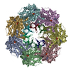
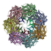
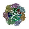
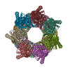
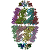
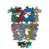
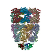
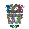
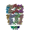
| 構造データ | EMDB-17418, PDB-8p4m: EMDB-17420, PDB-8p4n: EMDB-17421, PDB-8p4o:  EMDB-17422: Density for MetK encapsulated in the GroEL7-GroES7 cage  EMDB-17423: Symmetry-averaged GroEL7-GroES7 chamber with encapsulated disordered substrate MetK obtained by in vitro cryo electron tomography  EMDB-17424: Symmetry-averaged GroEL7-GroES7 chamber with encapsulated ordered substrate MetK obtained by in vitro cryo electron tomography EMDB-17425, PDB-8p4p: EMDB-17426, PDB-8p4r:  EMDB-17534: Cryo-EM structure of a D7-symmetrical GroEL14-GroES14 complex in presence of ADP-BeFx  EMDB-17535: Cryo-EM structure of a C7-symmetrical GroEL14-GroES7 complex in presence of ADP-BeFx  EMDB-17559: In situ structure average of GroEL7-GroES7 chamber with no or disordered substrate in Escherichia coli cytosol obtained by cryo electron tomography  EMDB-17560: In situ structure average of GroEL7-GroES7 chamber with encapsulated, ordered substrate in Escherichia coli cytosol obtained by cryo electron tomography  EMDB-17561: Cryo-ET subtomogram of 70S ribosomes in Escherichia coli cells at 37 and 46 degrees centigrade and in Escherichia coli cells overexpressing GroELS and MetK  EMDB-17562: Cryo-ET subtomogram of 70S ribosomes in Escherichia coli cells overexpressing GroEL  EMDB-17563: CryoEM structure of a GroEL14-GroES7 cage with encapsulated ordered substrate MetK in the presence of ADP-BeFx  EMDB-17564: CryoEM structure of a GroEL14-GroES7 cage with encapsulated disordered substrate MetK in the presence of ADP-BeFx  EMDB-17565: CryoEM structure of a GroEL14-GroES14 cage with two encapsulated disordered MetK substrates in the presence of ADP-BeFx  EMDB-17566: CryoEM structure of a (GroEL)14-(GroES)14 complex with encapsulated ordered MetK substrate in one chamber and no or disordered MetK substrate in the other chamber in the presence of ADP-BeFx  EMDB-17567: Conformer 1 of the (GroEL)14(GroES)14 complex with two encapsulated, ordered and near-native MetK substrate molecules in the presence of ADP-BeFx  EMDB-17568: Conformer 2 of the (GroEL)14(GroES)14 complex with two encapsulated, near-native and ordered MetK substrates in the presence of ADP-BeFx  EMDB-17569: Conformer 3 of the (GroEL)14(GroES)14 complex with two encapsulated, near-native and ordered MetK substrates in the presence of ADP-BeFx  EMDB-17570: Conformer 4 of the (GroEL)14(GroES)14 complex with two encapsulated, near-native and ordered MetK substrates in the presence of ADP-BeFx  EMDB-17571: Conformer 5 of the (GroEL)14(GroES)14 complex with two encapsulated, near-native and ordered MetK substrates in the presence of ADP-BeFx  EMDB-17572: Conformer 6 of the (GroEL)14(GroES)14 complex with two encapsulated, near-native and ordered MetK substrates in the presence of ADP-BeFx  EMDB-17573: Conformer 7 of the (GroEL)14(GroES)14 complex with two encapsulated, near-native and ordered MetK substrates in the presence of ADP-BeFx EMDB-18735, PDB-8qxs: EMDB-18736, PDB-8qxt: EMDB-18737, PDB-8qxu: EMDB-18738, PDB-8qxv: |
| 化合物 |  ChemComp-ADP:  ChemComp-MG:  ChemComp-BEF:  ChemComp-K:  ChemComp-HOH:  ChemComp-ATP: |
| 由来 |
|
 キーワード キーワード | CHAPERONE / Chaperonin / Folding cage / proteostasis / heat shock / ATPase |
 ムービー
ムービー コントローラー
 構造ビューア
 万見文献について


