 Movie
Movie Controller
 Structure viewers
 About Yorodumi Papers


+Search query
-Structure paper
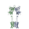
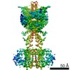
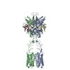
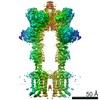
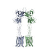
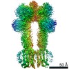
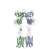
| Title | Structures of human mGlu2 and mGlu7 homo- and heterodimers. |
|---|---|
| Journal, issue, pages | Nature, Vol. 594, Issue 7864, Page 589-593, Year 2021 |
| Publish date | Jun 16, 2021 |
 Authors Authors | Juan Du / Dejian Wang / Hongcheng Fan / Chanjuan Xu / Linhua Tai / Shuling Lin / Shuo Han / Qiuxiang Tan / Xinwei Wang / Tuo Xu / Hui Zhang / Xiaojing Chu / Cuiying Yi / Peng Liu / Xiaomei Wang / Yu Zhou / Jean-Philippe Pin / Philippe Rondard / Hong Liu / Jianfeng Liu / Fei Sun / Beili Wu / Qiang Zhao /   |
| PubMed Abstract | The metabotropic glutamate receptors (mGlus) are involved in the modulation of synaptic transmission and neuronal excitability in the central nervous system. These receptors probably exist as both ...The metabotropic glutamate receptors (mGlus) are involved in the modulation of synaptic transmission and neuronal excitability in the central nervous system. These receptors probably exist as both homo- and heterodimers that have unique pharmacological and functional properties. Here we report four cryo-electron microscopy structures of the human mGlu subtypes mGlu2 and mGlu7, including inactive mGlu2 and mGlu7 homodimers; mGlu2 homodimer bound to an agonist and a positive allosteric modulator; and inactive mGlu2-mGlu7 heterodimer. We observed a subtype-dependent dimerization mode for these mGlus, as a unique dimer interface that is mediated by helix IV (and that is important for limiting receptor activity) exists only in the inactive mGlu2 structure. The structures provide molecular details of the inter- and intra-subunit conformational changes that are required for receptor activation, which distinguish class C G-protein-coupled receptors from those in classes A and B. Furthermore, our structure and functional studies of the mGlu2-mGlu7 heterodimer suggest that the mGlu7 subunit has a dominant role in controlling dimeric association and G-protein activation in the heterodimer. These insights into mGlu homo- and heterodimers highlight the complex landscape of mGlu dimerization and activation. |
 External links External links | Nature / PubMed:34135509 |
| Methods | EM (single particle) / X-ray diffraction |
| Resolution | 2.5 - 4.0 Å |
| Structure data | EMDB-31235, PDB-7epa: EMDB-31236, PDB-7epb: EMDB-31237, PDB-7epc: EMDB-31238, PDB-7epd: 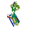 PDB-7epe: 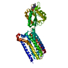 PDB-7epf: |
| Chemicals |  ChemComp-40F:  ChemComp-FMN: 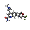 ChemComp-J9R:  ChemComp-J9U: |
| Source |
|
 Keywords Keywords | MEMBRANE PROTEIN / cryo-EM structure / GPCR |

 homo sapiens (human)
