 Movie
Movie Controller


+ Open data

- Basic information
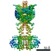
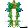
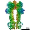
| Entry | Database: PDB / ID: 7epa | |||||||||||||||
|---|---|---|---|---|---|---|---|---|---|---|---|---|---|---|---|---|
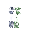
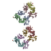
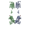
| Title | Cryo-EM structure of inactive mGlu2 homodimer | |||||||||||||||
 Components Components | Metabotropic glutamate receptor 2 | |||||||||||||||
 Keywords Keywords | MEMBRANE PROTEIN / cryo-EM structure / GPCR | |||||||||||||||
| Function / homology |  Function and homology information Function and homology informationregulation of response to drug / group II metabotropic glutamate receptor activity / intracellular glutamate homeostasis / behavioral response to nicotine / negative regulation of adenylate cyclase activity / G protein-coupled glutamate receptor signaling pathway / glutamate secretion / Class C/3 (Metabotropic glutamate/pheromone receptors) / glutamate receptor activity / long-term synaptic depression ...regulation of response to drug / group II metabotropic glutamate receptor activity / intracellular glutamate homeostasis / behavioral response to nicotine / negative regulation of adenylate cyclase activity / G protein-coupled glutamate receptor signaling pathway / glutamate secretion / Class C/3 (Metabotropic glutamate/pheromone receptors) / glutamate receptor activity / long-term synaptic depression / regulation of glutamate secretion / astrocyte projection / cellular response to stress / regulation of dopamine secretion / regulation of synaptic transmission, glutamatergic / presynaptic modulation of chemical synaptic transmission / response to cocaine / calcium channel regulator activity / G protein-coupled receptor activity / presynaptic membrane / scaffold protein binding / gene expression / G alpha (i) signalling events / chemical synaptic transmission / postsynaptic membrane / positive regulation of phosphatidylinositol 3-kinase/protein kinase B signal transduction / axon / dendrite / glutamatergic synapse / plasma membrane Similarity search - Function | |||||||||||||||
| Biological species |  Homo sapiens (human) Homo sapiens (human) | |||||||||||||||
| Method | ELECTRON MICROSCOPY / single particle reconstruction / cryo EM / Resolution: 3.6 Å | |||||||||||||||
 Authors Authors | Du, J. / Wang, D. / Fan, H. / Tai, L. / Lin, S. / Han, S. / Sun, F. / Wu, B. / Zhao, Q. | |||||||||||||||
| Funding support |  China, 4items China, 4items
| |||||||||||||||
 Citation Citation | Journal: Nature / Year: 2021 Title: Structures of human mGlu2 and mGlu7 homo- and heterodimers. Authors: Juan Du / Dejian Wang / Hongcheng Fan / Chanjuan Xu / Linhua Tai / Shuling Lin / Shuo Han / Qiuxiang Tan / Xinwei Wang / Tuo Xu / Hui Zhang / Xiaojing Chu / Cuiying Yi / Peng Liu / Xiaomei ...Authors: Juan Du / Dejian Wang / Hongcheng Fan / Chanjuan Xu / Linhua Tai / Shuling Lin / Shuo Han / Qiuxiang Tan / Xinwei Wang / Tuo Xu / Hui Zhang / Xiaojing Chu / Cuiying Yi / Peng Liu / Xiaomei Wang / Yu Zhou / Jean-Philippe Pin / Philippe Rondard / Hong Liu / Jianfeng Liu / Fei Sun / Beili Wu / Qiang Zhao /  Abstract: The metabotropic glutamate receptors (mGlus) are involved in the modulation of synaptic transmission and neuronal excitability in the central nervous system. These receptors probably exist as both ...The metabotropic glutamate receptors (mGlus) are involved in the modulation of synaptic transmission and neuronal excitability in the central nervous system. These receptors probably exist as both homo- and heterodimers that have unique pharmacological and functional properties. Here we report four cryo-electron microscopy structures of the human mGlu subtypes mGlu2 and mGlu7, including inactive mGlu2 and mGlu7 homodimers; mGlu2 homodimer bound to an agonist and a positive allosteric modulator; and inactive mGlu2-mGlu7 heterodimer. We observed a subtype-dependent dimerization mode for these mGlus, as a unique dimer interface that is mediated by helix IV (and that is important for limiting receptor activity) exists only in the inactive mGlu2 structure. The structures provide molecular details of the inter- and intra-subunit conformational changes that are required for receptor activation, which distinguish class C G-protein-coupled receptors from those in classes A and B. Furthermore, our structure and functional studies of the mGlu2-mGlu7 heterodimer suggest that the mGlu7 subunit has a dominant role in controlling dimeric association and G-protein activation in the heterodimer. These insights into mGlu homo- and heterodimers highlight the complex landscape of mGlu dimerization and activation. | |||||||||||||||
| History |
|
- Structure visualization
| Movie |
Movie viewer |
|---|---|
| Structure viewer | Molecule: MolmilJmol/JSmol |
- Downloads & links
-Download
| PDBx/mmCIF format | 7epa.cif.gz | 317.2 KB | Display | PDBx/mmCIF format |
|---|---|---|---|---|
| PDB format | pdb7epa.ent.gz | 233 KB | Display | PDB format |
| PDBx/mmJSON format | 7epa.json.gz | Tree view | PDBx/mmJSON format | |
| Others |  Other downloads Other downloads |
-Validation report
| Arichive directory | https://data.pdbj.org/pub/pdb/validation_reports/ep/7epaftp://data.pdbj.org/pub/pdb/validation_reports/ep/7epa | HTTPS FTP |
|---|
-Related structure data
| Related structure data |  31235MC 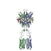 7epbC 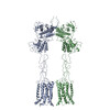 7epcC 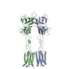 7epdC 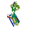 7epeC 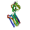 7epfC M: map data used to model this data C: citing same article ( |
|---|---|
| Similar structure data |


-Links
 PDBj
PDBj

- Assembly
| Deposited unit | 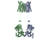
|
|---|---|
| 1 |
|
-Components
| #1: Protein | Mass: 91111.289 Da / Num. of mol.: 2 / Mutation: N655Y, H815Y Source method: isolated from a genetically manipulated source Source: (gene. exp.) Homo sapiens (human) / Gene: GRM2, GPRC1B, MGLUR2Production host:  References: UniProt: Q14416 Has protein modification | Y | |
|---|
-Experimental details
-Experiment
| Experiment | Method: ELECTRON MICROSCOPY |
|---|---|
| EM experiment | Aggregation state: PARTICLE / 3D reconstruction method: single particle reconstruction |
- Sample preparation
| Component | Name: Inactive mGlu2 homodimer / Type: COMPLEX / Entity ID: all / Source: RECOMBINANT |
|---|---|
| Source (natural) | Organism: Homo sapiens (human) |
| Source (recombinant) | Organism: |
| Buffer solution | pH: 7.5 |
| Specimen | Embedding applied: NO / Shadowing applied: NO / Staining applied: NO / Vitrification applied: YES |
| Vitrification | Cryogen name: ETHANE |
- Electron microscopy imaging
| Experimental equipment |  Model: Titan Krios / Image courtesy: FEI Company |
|---|---|
| Microscopy | Model: FEI TITAN KRIOS |
| Electron gun | Electron source:  FIELD EMISSION GUN / Accelerating voltage: 300 kV / Illumination mode: SPOT SCAN FIELD EMISSION GUN / Accelerating voltage: 300 kV / Illumination mode: SPOT SCAN |
| Electron lens | Mode: BRIGHT FIELD |
| Image recording | Electron dose: 1.75 e/Å2 / Film or detector model: GATAN K2 QUANTUM (4k x 4k) |
- Processing
| CTF correction | Type: NONE | ||||||||||||||||||||||||
|---|---|---|---|---|---|---|---|---|---|---|---|---|---|---|---|---|---|---|---|---|---|---|---|---|---|
| 3D reconstruction | Resolution: 3.6 Å / Resolution method: FSC 0.143 CUT-OFF / Num. of particles: 257133 / Symmetry type: POINT | ||||||||||||||||||||||||
| Refinement | Stereochemistry target values: GeoStd + Monomer Library + CDL v1.2 | ||||||||||||||||||||||||
| Displacement parameters | Biso mean: 141.92 Å2 | ||||||||||||||||||||||||
| Refine LS restraints |
|
