 Movie
Movie Controller


+ Open data

- Basic information
| Entry | Database: EMDB / ID: EMD-31237 | |||||||||||||||
|---|---|---|---|---|---|---|---|---|---|---|---|---|---|---|---|---|
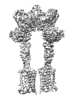
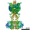
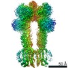
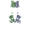
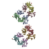
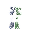
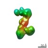
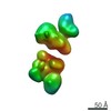
| Title | Cryo-EM structure of inactive mGlu7 homodimer | |||||||||||||||
 Map data Map data | ||||||||||||||||
 Sample Sample |
| |||||||||||||||
 Keywords Keywords | Cryo-EM structure / membrane protein / GPCR | |||||||||||||||
| Function / homology | Isoform 3 of Metabotropic glutamate receptor 7 Function and homology information Function and homology information | |||||||||||||||
| Biological species |  Homo sapiens (human) Homo sapiens (human) | |||||||||||||||
| Method | single particle reconstruction / cryo EM / Resolution: 4.0 Å | |||||||||||||||
 Authors Authors | Du J / Wang D | |||||||||||||||
| Funding support |  China, 4 items China, 4 items
| |||||||||||||||
 Citation Citation | Journal: Nature / Year: 2021 Title: Structures of human mGlu2 and mGlu7 homo- and heterodimers. Authors: Juan Du / Dejian Wang / Hongcheng Fan / Chanjuan Xu / Linhua Tai / Shuling Lin / Shuo Han / Qiuxiang Tan / Xinwei Wang / Tuo Xu / Hui Zhang / Xiaojing Chu / Cuiying Yi / Peng Liu / Xiaomei ...Authors: Juan Du / Dejian Wang / Hongcheng Fan / Chanjuan Xu / Linhua Tai / Shuling Lin / Shuo Han / Qiuxiang Tan / Xinwei Wang / Tuo Xu / Hui Zhang / Xiaojing Chu / Cuiying Yi / Peng Liu / Xiaomei Wang / Yu Zhou / Jean-Philippe Pin / Philippe Rondard / Hong Liu / Jianfeng Liu / Fei Sun / Beili Wu / Qiang Zhao /  Abstract: The metabotropic glutamate receptors (mGlus) are involved in the modulation of synaptic transmission and neuronal excitability in the central nervous system. These receptors probably exist as both ...The metabotropic glutamate receptors (mGlus) are involved in the modulation of synaptic transmission and neuronal excitability in the central nervous system. These receptors probably exist as both homo- and heterodimers that have unique pharmacological and functional properties. Here we report four cryo-electron microscopy structures of the human mGlu subtypes mGlu2 and mGlu7, including inactive mGlu2 and mGlu7 homodimers; mGlu2 homodimer bound to an agonist and a positive allosteric modulator; and inactive mGlu2-mGlu7 heterodimer. We observed a subtype-dependent dimerization mode for these mGlus, as a unique dimer interface that is mediated by helix IV (and that is important for limiting receptor activity) exists only in the inactive mGlu2 structure. The structures provide molecular details of the inter- and intra-subunit conformational changes that are required for receptor activation, which distinguish class C G-protein-coupled receptors from those in classes A and B. Furthermore, our structure and functional studies of the mGlu2-mGlu7 heterodimer suggest that the mGlu7 subunit has a dominant role in controlling dimeric association and G-protein activation in the heterodimer. These insights into mGlu homo- and heterodimers highlight the complex landscape of mGlu dimerization and activation. | |||||||||||||||
| History |
|
- Structure visualization
| Movie |
Movie viewer |
|---|---|
| Structure viewer | EM map: SurfViewMolmilJmol/JSmol |
| Supplemental images |
- Downloads & links
-EMDB archive
| Map data | emd_31237.map.gz | 147.1 MB | EMDB map data format | |
|---|---|---|---|---|
| Header (meta data) | emd-31237-v30.xmlemd-31237.xml | 10.9 KB 10.9 KB | Display Display | EMDB header |
| Images |  emd_31237.png emd_31237.png | 101.6 KB | ||
| Filedesc metadata | emd-31237.cif.gz | 5.4 KB | ||
| Archive directory |  http://ftp.pdbj.org/pub/emdb/structures/EMD-31237ftp://ftp.pdbj.org/pub/emdb/structures/EMD-31237 http://ftp.pdbj.org/pub/emdb/structures/EMD-31237ftp://ftp.pdbj.org/pub/emdb/structures/EMD-31237 | HTTPS FTP |
-Related structure data
| Related structure data |  7epcMC  7epaC  7epbC 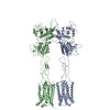 7epdC 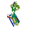 7epeC 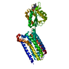 7epfC M: atomic model generated by this map C: citing same article ( |
|---|---|
| Similar structure data |


-Links
| EMDB pages | EMDB (EBI/PDBe) / EMDataResource |
|---|
-Map
| File | Download / File: emd_31237.map.gz / Format: CCP4 / Size: 181 MB / Type: IMAGE STORED AS FLOATING POINT NUMBER (4 BYTES) | ||||||||||||||||||||||||||||||||||||||||||||||||||||||||||||||||||||
|---|---|---|---|---|---|---|---|---|---|---|---|---|---|---|---|---|---|---|---|---|---|---|---|---|---|---|---|---|---|---|---|---|---|---|---|---|---|---|---|---|---|---|---|---|---|---|---|---|---|---|---|---|---|---|---|---|---|---|---|---|---|---|---|---|---|---|---|---|---|
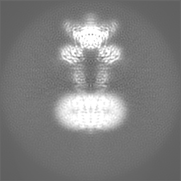
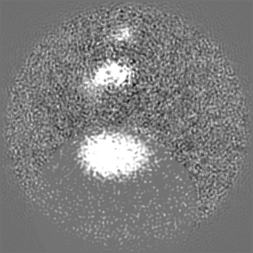
| Projections & slices | Image control
Images are generated by Spider. | ||||||||||||||||||||||||||||||||||||||||||||||||||||||||||||||||||||
| Voxel size | X=Y=Z: 0.8 Å | ||||||||||||||||||||||||||||||||||||||||||||||||||||||||||||||||||||
| Density |
| ||||||||||||||||||||||||||||||||||||||||||||||||||||||||||||||||||||
| Symmetry | Space group: 1 | ||||||||||||||||||||||||||||||||||||||||||||||||||||||||||||||||||||
| Details | EMDB XML:
CCP4 map header:
| ||||||||||||||||||||||||||||||||||||||||||||||||||||||||||||||||||||
 Z (Sec.)
 Y (Row.)
 X (Col.)


-Supplemental data
- Sample components
-Entire : Inactive mGlu7 homodimer
| Entire | Name: Inactive mGlu7 homodimer |
|---|---|
| Components |
|
-Supramolecule #1: Inactive mGlu7 homodimer
| Supramolecule | Name: Inactive mGlu7 homodimer / type: complex / ID: 1 / Parent: 0 / Macromolecule list: all |
|---|---|
| Source (natural) | Organism: Homo sapiens (human) |
-Macromolecule #1: Isoform 3 of Metabotropic glutamate receptor 7
| Macromolecule | Name: Isoform 3 of Metabotropic glutamate receptor 7 / type: protein_or_peptide / ID: 1 / Number of copies: 2 / Enantiomer: LEVO |
|---|---|
| Source (natural) | Organism: Homo sapiens (human) |
| Molecular weight | Theoretical: 96.276977 KDa |
| Recombinant expression | Organism:  |
| Sequence | String: GAPQEMYAPH SIRIEGDVTL GGLFPVHAKG PSGVPCGDIK RENGIHRLEA MLYALDQINS DPNLLPNVTL GARILDTCSR DTYALEQSL TFVQALIQKD TSDVRCTNGE PPVFVKPEKV VGVIGASGSS VSIMVANILR LFQIPQISYA STAPELSDDR R YDFFSRVV ...String: GAPQEMYAPH SIRIEGDVTL GGLFPVHAKG PSGVPCGDIK RENGIHRLEA MLYALDQINS DPNLLPNVTL GARILDTCSR DTYALEQSL TFVQALIQKD TSDVRCTNGE PPVFVKPEKV VGVIGASGSS VSIMVANILR LFQIPQISYA STAPELSDDR R YDFFSRVV PPDSFQAQAM VDIVKALGWN YVSTLASEGS YGEKGVESFT QISKEAGGLC IAQSVRIPQE RKDRTIDFDR II KQLLDTP NSRAVVIFAN DEDIKQILAA AKRADQVGHF LWVGSDSWGS KINPLHQHED IAEGAITIQP KRATVEGFDA YFT SRTLEN NRRNVWFAEY WEENFNCKLT ISGSKKEDTD RKCTGQERIG KDSNYEQEGK VQFVIDAVYA MAHALHHMNK DLCA DYRGV CPEMEQAGGK KLLKYIRNVN FNGSAGTPVM FNKNGDAPGR YDIFQYQTTN TSNPGYRLIG QWTDELQLNI EDMQW GKGV REIPASVCTL PCKPGQRKKT QKGTPCCWTC EPCDGYQYQF DEMTCQHCPY DQRPNENRTG CQDIPIIKLE WHSPWA VIP VFLAMLGIIA TIFVMATFIR YNDTPIVRAS GRELSYVLLT GIFLCYIITF LMIAKPDVAV CSFRRVFLGL GMCISYA AL LTKTYRIYRI FEQGKKSVTA PRLISPTSQL AITSSLISVQ LLGVFIWFIV DPPNIIIDYD EHKTMNPEQA RGVLKCDI T DLQIICSLGY SILLMVTCTV YAFKTRGVPE NFNEAKYIGF TMYTTCIVWL AFIPIFFGTA QSAEKLYIQT TTLTISMNL SASVALGMLY MPKVYIIIFH PELNVQKREF LEVLFQGPGS GSWSHPQFEK DYKDDDD UniProtKB: Isoform 3 of Metabotropic glutamate receptor 7 |
-Experimental details
-Structure determination
| Method | cryo EM |
|---|---|
 Processing Processing | single particle reconstruction |
| Aggregation state | particle |
-Sample preparation
| Buffer | pH: 7.5 |
|---|---|
| Vitrification | Cryogen name: ETHANE |
- Electron microscopy
| Microscope | FEI TALOS ARCTICA |
|---|---|
| Image recording | Film or detector model: GATAN K2 QUANTUM (4k x 4k) / Average electron dose: 1.47 e/Å2 |
| Electron beam | Acceleration voltage: 200 kV / Electron source:  FIELD EMISSION GUN FIELD EMISSION GUN |
| Electron optics | Illumination mode: SPOT SCAN / Imaging mode: BRIGHT FIELD |
| Experimental equipment |  Model: Talos Arctica / Image courtesy: FEI Company |
-Image processing
| Startup model | Type of model: INSILICO MODEL |
|---|---|
| Final reconstruction | Resolution.type: BY AUTHOR / Resolution: 4.0 Å / Resolution method: FSC 0.143 CUT-OFF / Number images used: 1011214 |
| Initial angle assignment | Type: MAXIMUM LIKELIHOOD |
| Final angle assignment | Type: MAXIMUM LIKELIHOOD |
