 Movie
Movie Controller


+ Open data

- Basic information
| Entry | Database: EMDB / ID: EMD-31238 | |||||||||||||||
|---|---|---|---|---|---|---|---|---|---|---|---|---|---|---|---|---|
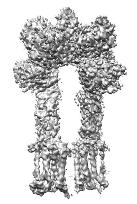
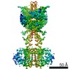
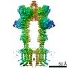
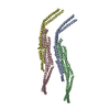
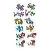
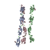
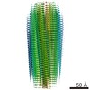
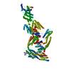
| Title | Cryo-EM structure of inactive mGlu2-7 heterodimer | |||||||||||||||
 Map data Map data | ||||||||||||||||
 Sample Sample |
| |||||||||||||||
 Keywords Keywords | Cryo-EM structure / membrane protein / GPCR | |||||||||||||||
| Function / homology |  Function and homology information Function and homology informationregulation of response to drug / group II metabotropic glutamate receptor activity / intracellular glutamate homeostasis / behavioral response to nicotine / negative regulation of adenylate cyclase activity / G protein-coupled glutamate receptor signaling pathway / glutamate secretion / Class C/3 (Metabotropic glutamate/pheromone receptors) / glutamate receptor activity / long-term synaptic depression ...regulation of response to drug / group II metabotropic glutamate receptor activity / intracellular glutamate homeostasis / behavioral response to nicotine / negative regulation of adenylate cyclase activity / G protein-coupled glutamate receptor signaling pathway / glutamate secretion / Class C/3 (Metabotropic glutamate/pheromone receptors) / glutamate receptor activity / long-term synaptic depression / regulation of glutamate secretion / astrocyte projection / cellular response to stress / regulation of dopamine secretion / regulation of synaptic transmission, glutamatergic / presynaptic modulation of chemical synaptic transmission / peptidylprolyl isomerase / peptidyl-prolyl cis-trans isomerase activity / response to cocaine / calcium channel regulator activity / G protein-coupled receptor activity / presynaptic membrane / scaffold protein binding / gene expression / G alpha (i) signalling events / chemical synaptic transmission / postsynaptic membrane / positive regulation of phosphatidylinositol 3-kinase/protein kinase B signal transduction / axon / dendrite / glutamatergic synapse / plasma membrane Similarity search - Function | |||||||||||||||
| Biological species |  Homo sapiens (human) Homo sapiens (human) | |||||||||||||||
| Method | single particle reconstruction / cryo EM / Resolution: 3.9 Å | |||||||||||||||
 Authors Authors | Du J / Wang D | |||||||||||||||
| Funding support |  China, 4 items China, 4 items
| |||||||||||||||
 Citation Citation | Journal: Nature / Year: 2021 Title: Structures of human mGlu2 and mGlu7 homo- and heterodimers. Authors: Juan Du / Dejian Wang / Hongcheng Fan / Chanjuan Xu / Linhua Tai / Shuling Lin / Shuo Han / Qiuxiang Tan / Xinwei Wang / Tuo Xu / Hui Zhang / Xiaojing Chu / Cuiying Yi / Peng Liu / Xiaomei ...Authors: Juan Du / Dejian Wang / Hongcheng Fan / Chanjuan Xu / Linhua Tai / Shuling Lin / Shuo Han / Qiuxiang Tan / Xinwei Wang / Tuo Xu / Hui Zhang / Xiaojing Chu / Cuiying Yi / Peng Liu / Xiaomei Wang / Yu Zhou / Jean-Philippe Pin / Philippe Rondard / Hong Liu / Jianfeng Liu / Fei Sun / Beili Wu / Qiang Zhao /  Abstract: The metabotropic glutamate receptors (mGlus) are involved in the modulation of synaptic transmission and neuronal excitability in the central nervous system. These receptors probably exist as both ...The metabotropic glutamate receptors (mGlus) are involved in the modulation of synaptic transmission and neuronal excitability in the central nervous system. These receptors probably exist as both homo- and heterodimers that have unique pharmacological and functional properties. Here we report four cryo-electron microscopy structures of the human mGlu subtypes mGlu2 and mGlu7, including inactive mGlu2 and mGlu7 homodimers; mGlu2 homodimer bound to an agonist and a positive allosteric modulator; and inactive mGlu2-mGlu7 heterodimer. We observed a subtype-dependent dimerization mode for these mGlus, as a unique dimer interface that is mediated by helix IV (and that is important for limiting receptor activity) exists only in the inactive mGlu2 structure. The structures provide molecular details of the inter- and intra-subunit conformational changes that are required for receptor activation, which distinguish class C G-protein-coupled receptors from those in classes A and B. Furthermore, our structure and functional studies of the mGlu2-mGlu7 heterodimer suggest that the mGlu7 subunit has a dominant role in controlling dimeric association and G-protein activation in the heterodimer. These insights into mGlu homo- and heterodimers highlight the complex landscape of mGlu dimerization and activation. | |||||||||||||||
| History |
|
- Structure visualization
| Movie |
Movie viewer |
|---|---|
| Structure viewer | EM map: SurfViewMolmilJmol/JSmol |
| Supplemental images |
- Downloads & links
-EMDB archive
| Map data | emd_31238.map.gz | 95.9 MB | EMDB map data format | |
|---|---|---|---|---|
| Header (meta data) | emd-31238-v30.xmlemd-31238.xml | 13.1 KB 13.1 KB | Display Display | EMDB header |
| Images |  emd_31238.png emd_31238.png | 109.8 KB | ||
| Filedesc metadata | emd-31238.cif.gz | 6.2 KB | ||
| Archive directory |  http://ftp.pdbj.org/pub/emdb/structures/EMD-31238ftp://ftp.pdbj.org/pub/emdb/structures/EMD-31238 http://ftp.pdbj.org/pub/emdb/structures/EMD-31238ftp://ftp.pdbj.org/pub/emdb/structures/EMD-31238 | HTTPS FTP |
-Related structure data
| Related structure data |  7epdMC  7epaC  7epbC 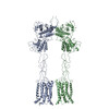 7epcC 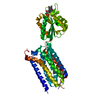 7epeC 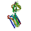 7epfC M: atomic model generated by this map C: citing same article ( |
|---|---|
| Similar structure data |


-Links
| EMDB pages | EMDB (EBI/PDBe) / EMDataResource |
|---|---|
| Related items in Molecule of the Month |


-Map
| File | Download / File: emd_31238.map.gz / Format: CCP4 / Size: 110 MB / Type: IMAGE STORED AS FLOATING POINT NUMBER (4 BYTES) | ||||||||||||||||||||||||||||||||||||||||||||||||||||||||||||||||||||
|---|---|---|---|---|---|---|---|---|---|---|---|---|---|---|---|---|---|---|---|---|---|---|---|---|---|---|---|---|---|---|---|---|---|---|---|---|---|---|---|---|---|---|---|---|---|---|---|---|---|---|---|---|---|---|---|---|---|---|---|---|---|---|---|---|---|---|---|---|---|
| Projections & slices | Image control
Images are generated by Spider. generated in cubic-lattice coordinate | ||||||||||||||||||||||||||||||||||||||||||||||||||||||||||||||||||||
| Voxel size | X=Y=Z: 1.045 Å | ||||||||||||||||||||||||||||||||||||||||||||||||||||||||||||||||||||
| Density |
| ||||||||||||||||||||||||||||||||||||||||||||||||||||||||||||||||||||
| Symmetry | Space group: 1 | ||||||||||||||||||||||||||||||||||||||||||||||||||||||||||||||||||||
| Details | EMDB XML:
CCP4 map header:
| ||||||||||||||||||||||||||||||||||||||||||||||||||||||||||||||||||||
 Z (Sec.)
 Y (Row.)
 X (Col.)


-Supplemental data
- Sample components
-Entire : Inactive mGlu2-7 heterodimer
| Entire | Name: Inactive mGlu2-7 heterodimer |
|---|---|
| Components |
|
-Supramolecule #1: Inactive mGlu2-7 heterodimer
| Supramolecule | Name: Inactive mGlu2-7 heterodimer / type: complex / ID: 1 / Parent: 0 / Macromolecule list: all |
|---|---|
| Source (natural) | Organism: Homo sapiens (human) |
-Macromolecule #1: Metabotropic glutamate receptor 2,Peptidylprolyl isomerase
| Macromolecule | Name: Metabotropic glutamate receptor 2,Peptidylprolyl isomerase type: protein_or_peptide / ID: 1 / Number of copies: 1 / Enantiomer: LEVO / EC number: peptidylprolyl isomerase |
|---|---|
| Source (natural) | Organism: Homo sapiens (human) |
| Molecular weight | Theoretical: 104.904 KDa |
| Recombinant expression | Organism:  |
| Sequence | String: DYKDDDDGAP EGPAKKVLTL EGDLVLGGLF PVHQKGGPAE DCGPVNEHRG IQRLEAMLFA LDRINRDPHL LPGVRLGAHI LDSCSKDTH ALEQALDFVR ASLSRGADGS RHICPDGSYA THGDAPTAIT GVIGGSYSDV SIQVANLLRL FQIPQISYAS T SAKLSDKS ...String: DYKDDDDGAP EGPAKKVLTL EGDLVLGGLF PVHQKGGPAE DCGPVNEHRG IQRLEAMLFA LDRINRDPHL LPGVRLGAHI LDSCSKDTH ALEQALDFVR ASLSRGADGS RHICPDGSYA THGDAPTAIT GVIGGSYSDV SIQVANLLRL FQIPQISYAS T SAKLSDKS RYDYFARTVP PDFFQAKAMA EILRFFNWTY VSTVASEGDY GETGIEAFEL EARARNICVA TSEKVGRAMS RA AFEGVVR ALLQKPSARV AVLFTRSEDA RELLAASQRL NASFTWVASD GWGALESVVA GSEGAAEGAI TIELASYPIS DFA SYFQSL DPWNNSRNPW FREFWEQRFR CSFRQRDCAA HSLRAVPFEQ ESKIMFVVNA VYAMAHALHN MHRALCPNTT RLCD AMRPV NGRRLYKDFV LNVKFDAPFR PADTHNEVRF DRFGDGIGRY NIFTYLRAGS GRYRYQKVGY WAEGLTLDTS LIPWA SPSA GPLPASRCSE PCLQNEVKSV QPGEVCCWLC IPCQPYEYRL DEFTCADCGL GYWPNASLTG CFELPQEYIR WGDAWA VGP VTIACLGALA TLFVLGVFVR HNATPVVKAS GRELCYILLG GVFLCYCMTF IFIAKPSTAV CTLRRLGLGT AFSVCYS AL LTKTYRIARI FGGAREGAQR PRFISPASQV AICLALISGQ LLIVVAWLVV EAPGTGKETA PERREVVTLR CNHRDASM L GSLAYNVLLI ALCTLYAFKT RKCPENFNEA KFIGFTMYTT CIIWLAFLPI FYVTSSDYRV QTTTMCVSVS LSGSVVLGC LFAPKLYIIL FQPQKNVAAA GVQVETISPG DGRTFPKRGQ TCVVHYTGML EDGKKFDSSR DRNKPFKFML GKQEVIRGWE EGVAQMSVG QRAKLTISPD YAYGATGHPG IIPPHATLVF DVELLKLEEF LEVLFQGPHH HHHHHHHH UniProtKB: Metabotropic glutamate receptor 2, peptidylprolyl isomerase |
-Macromolecule #2: Isoform 3 of Metabotropic glutamate receptor 7
| Macromolecule | Name: Isoform 3 of Metabotropic glutamate receptor 7 / type: protein_or_peptide / ID: 2 / Number of copies: 1 / Enantiomer: LEVO |
|---|---|
| Source (natural) | Organism: Homo sapiens (human) |
| Molecular weight | Theoretical: 109.243688 KDa |
| Recombinant expression | Organism: |
| Sequence | String: GSWSHPQFEK GSGSWSHPQF EKGSLEVLFQ GPGAPQEMYA PHSIRIEGDV TLGGLFPVHA KGPSGVPCGD IKRENGIHRL EAMLYALDQ INSDPNLLPN VTLGARILDT CSRDTYALEQ SLTFVQALIQ KDTSDVRCTN GEPPVFVKPE KVVGVIGASG S SVSIMVAN ...String: GSWSHPQFEK GSGSWSHPQF EKGSLEVLFQ GPGAPQEMYA PHSIRIEGDV TLGGLFPVHA KGPSGVPCGD IKRENGIHRL EAMLYALDQ INSDPNLLPN VTLGARILDT CSRDTYALEQ SLTFVQALIQ KDTSDVRCTN GEPPVFVKPE KVVGVIGASG S SVSIMVAN ILRLFQIPQI SYASTAPELS DDRRYDFFSR VVPPDSFQAQ AMVDIVKALG WNYVSTLASE GSYGEKGVES FT QISKEAG GLCIAQSVRI PQERKDRTID FDRIIKQLLD TPNSRAVVIF ANDEDIKQIL AAAKRADQVG HFLWVGSDSW GSK INPLHQ HEDIAEGAIT IQPKRATVEG FDAYFTSRTL ENNRRNVWFA EYWEENFNCK LTISGSKKED TDRKCTGQER IGKD SNYEQ EGKVQFVIDA VYAMAHALHH MNKDLCADYR GVCPEMEQAG GKKLLKYIRN VNFNGSAGTP VMFNKNGDAP GRYDI FQYQ TTNTSNPGYR LIGQWTDELQ LNIEDMQWGK GVREIPASVC TLPCKPGQRK KTQKGTPCCW TCEPCDGYQY QFDEMT CQH CPYDQRPNEN RTGCQDIPII KLEWHSPWAV IPVFLAMLGI IATIFVMATF IRYNDTPIVR ASGRELSYVL LTGIFLC YI ITFLMIAKPD VAVCSFRRVF LGLGMCISYA ALLTKTNRIY RIFEQGKKSV TAPRLISPTS QLAITSSLIS VQLLGVFI W FIVDPPNIII DYDEHKTMNP EQARGVLKCD ITDLQIICSL GYSILLMVTC TVYAFKTRGV PENFNEAKYI GFTMYTTCI VWLAFIPIFF GTAQSAEKLY IQTTTLTISM NLSASVALGM LYMPKVYIII FHPELNVQKR AAAAILWHEM WHEGLEEASR LYFGERNVK GMFEVLEPLH AMMERGPQTL KETSFNQAYG RDLMEAQEWC RKYMKSGNVK DLTQAWDLYY HVFRRISKQE F DYKDDDD |
-Experimental details
-Structure determination
| Method | cryo EM |
|---|---|
 Processing Processing | single particle reconstruction |
| Aggregation state | particle |
-Sample preparation
| Buffer | pH: 7.5 |
|---|---|
| Vitrification | Cryogen name: ETHANE |
- Electron microscopy
| Microscope | FEI TITAN KRIOS |
|---|---|
| Image recording | Film or detector model: GATAN K3 BIOQUANTUM (6k x 4k) / Average electron dose: 2.1875 e/Å2 |
| Electron beam | Acceleration voltage: 300 kV / Electron source:  FIELD EMISSION GUN FIELD EMISSION GUN |
| Electron optics | Illumination mode: SPOT SCAN / Imaging mode: BRIGHT FIELD |
| Experimental equipment |  Model: Titan Krios / Image courtesy: FEI Company |
-Image processing
| Startup model | Type of model: INSILICO MODEL |
|---|---|
| Final reconstruction | Resolution.type: BY AUTHOR / Resolution: 3.9 Å / Resolution method: FSC 0.143 CUT-OFF / Number images used: 1113538 |
| Initial angle assignment | Type: MAXIMUM LIKELIHOOD |
| Final angle assignment | Type: MAXIMUM LIKELIHOOD |
