[English] 日本語
Yorodumi Papers
- Database of articles cited by EMDB/PDB/SASBDB data -

+
Search query

Keywords
Structure methods
Author
Journal
IF

-
Structure paper

TitleEbola and Marburg virus matrix layers are locally ordered assemblies of VP40 dimers.
Journal, issue, pagesElife, Vol. 9, Year 2020
Publish dateOct 5, 2020
AuthorsWilliam Wan / Mairi Clarke / Michael J Norris / Larissa Kolesnikova / Alexander Koehler / Zachary A Bornholdt / Stephan Becker / Erica Ollmann Saphire / John Ag Briggs /
PubMed AbstractFiloviruses such as Ebola and Marburg virus bud from the host membrane as enveloped virions. This process is achieved by the matrix protein VP40. When expressed alone, VP40 induces budding of ...Filoviruses such as Ebola and Marburg virus bud from the host membrane as enveloped virions. This process is achieved by the matrix protein VP40. When expressed alone, VP40 induces budding of filamentous virus-like particles, suggesting that localization to the plasma membrane, oligomerization into a matrix layer, and generation of membrane curvature are intrinsic properties of VP40. There has been no direct information on the structure of VP40 matrix layers within viruses or virus-like particles. We present structures of Ebola and Marburg VP40 matrix layers in intact virus-like particles, and within intact Marburg viruses. VP40 dimers assemble extended chains via C-terminal domain interactions. These chains stack to form 2D matrix lattices below the membrane surface. These lattices form a patchwork assembly across the membrane and suggesting that assembly may begin at multiple points. Our observations define the structure and arrangement of the matrix protein layer that mediates formation of filovirus particles.
External linksElife / PubMed:33016878 / PubMed Central
MethodsEM (subtomogram averaging) / X-ray diffraction
Resolution2.46 - 12.8 Å
Structure data

EMDB-11660:
Structure of VP40 matrix layer in EBOV NP-VP24-VP35-VP40 VLPs
Method: EM (subtomogram averaging) / Resolution: 10.2 Å

EMDB-11661:
Structure of VP40 matrix layer in EBOV VP40 VLPs
Method: EM (subtomogram averaging) / Resolution: 9.8 Å

EMDB-11662:
Structure of VP40 matrix layer in EBOV VP40-GP VLPs
Method: EM (subtomogram averaging) / Resolution: 9.9 Å

EMDB-11663:
Structure of VP40 matrix layer in intact MARV virions
Method: EM (subtomogram averaging) / Resolution: 9.6 Å

EMDB-11664:
Structure of VP40 matrix layer in MARV VP40 VLPs
Method: EM (subtomogram averaging) / Resolution: 10.8 Å

EMDB-11665:
Structure of GP in EBOV VP40-GP VLPs
Method: EM (subtomogram averaging) / Resolution: 12.8 Å

PDB-7jzj:
Crystal structure demonstrating CTD-CTD interactions of Zaire Ebola virus VP40 dimer
Method: X-RAY DIFFRACTION / Resolution: 2.46 Å

PDB-7jzt:
Low resolution crystal structure of Zaire Ebola virus VP40 in space group P6422
Method: X-RAY DIFFRACTION / Resolution: 3.77 Å

Chemicals

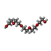
ChemComp-1PE:
PENTAETHYLENE GLYCOL / precipitant*YM

ChemComp-HOH:
WATER

Source
  • Ebola virus - Mayinga
  • Marburg virus - Musoke
  • zaire ebolavirus (strain mayinga-76)
KeywordsVIRAL PROTEIN / VP40 / matrix protein / Ebola virus

+
About Yorodumi Papers

-
News

-
Feb 9, 2022. New format data for meta-information of EMDB entries

New format data for meta-information of EMDB entries

  • Version 3 of the EMDB header file is now the official format.
  • The previous official version 1.9 will be removed from the archive.

Related info.:EMDB header

External links:wwPDB to switch to version 3 of the EMDB data model

-
Aug 12, 2020. Covid-19 info

Covid-19 info

URL: https://pdbj.org/emnavi/covid19.php

New page: Covid-19 featured information page in EM Navigator.

Related info.:Covid-19 info / Mar 5, 2020. Novel coronavirus structure data

+
Mar 5, 2020. Novel coronavirus structure data

Novel coronavirus structure data

Related info.:Yorodumi Speices / Aug 12, 2020. Covid-19 info

External links:COVID-19 featured content - PDBj / Molecule of the Month (242):Coronavirus Proteases

+
Jan 31, 2019. EMDB accession codes are about to change! (news from PDBe EMDB page)

EMDB accession codes are about to change! (news from PDBe EMDB page)

  • The allocation of 4 digits for EMDB accession codes will soon come to an end. Whilst these codes will remain in use, new EMDB accession codes will include an additional digit and will expand incrementally as the available range of codes is exhausted. The current 4-digit format prefixed with “EMD-” (i.e. EMD-XXXX) will advance to a 5-digit format (i.e. EMD-XXXXX), and so on. It is currently estimated that the 4-digit codes will be depleted around Spring 2019, at which point the 5-digit format will come into force.
  • The EM Navigator/Yorodumi systems omit the EMD- prefix.

Related info.:Q: What is EMD? / ID/Accession-code notation in Yorodumi/EM Navigator

External links:EMDB Accession Codes are Changing Soon! / Contact to PDBj

+
Jul 12, 2017. Major update of PDB

Major update of PDB

  • wwPDB released updated PDB data conforming to the new PDBx/mmCIF dictionary.
  • This is a major update changing the version number from 4 to 5, and with Remediation, in which all the entries are updated.
  • In this update, many items about electron microscopy experimental information are reorganized (e.g. em_software).
  • Now, EM Navigator and Yorodumi are based on the updated data.

External links:wwPDB Remediation / Enriched Model Files Conforming to OneDep Data Standards Now Available in the PDB FTP Archive

-
Yorodumi Papers

Database of articles cited by EMDB/PDB/SASBDB data

  • Database of articles cited by EMDB, PDB, and SASBDB entries
  • Using PubMed data

Related info.:EMDB / PDB / SASBDB / Yorodumi / EMN Papers / Changes in new EM Navigator and Yorodumi

Read more