 Movie
Movie Controller
 Structure viewers
 About Yorodumi Papers


+Search query
-Structure paper
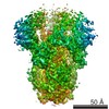
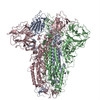
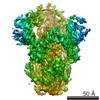
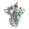
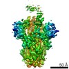
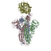
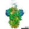
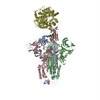
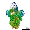
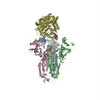
| Title | Cryo-EM structure of the SARS coronavirus spike glycoprotein in complex with its host cell receptor ACE2. |
|---|---|
| Journal, issue, pages | PLoS Pathog, Vol. 14, Issue 8, Page e1007236, Year 2018 |
| Publish date | Aug 13, 2018 |
 Authors Authors | Wenfei Song / Miao Gui / Xinquan Wang / Ye Xiang /  |
| PubMed Abstract | The trimeric SARS coronavirus (SARS-CoV) surface spike (S) glycoprotein consisting of three S1-S2 heterodimers binds the cellular receptor angiotensin-converting enzyme 2 (ACE2) and mediates fusion ...The trimeric SARS coronavirus (SARS-CoV) surface spike (S) glycoprotein consisting of three S1-S2 heterodimers binds the cellular receptor angiotensin-converting enzyme 2 (ACE2) and mediates fusion of the viral and cellular membranes through a pre- to postfusion conformation transition. Here, we report the structure of the SARS-CoV S glycoprotein in complex with its host cell receptor ACE2 revealed by cryo-electron microscopy (cryo-EM). The complex structure shows that only one receptor-binding domain of the trimeric S glycoprotein binds ACE2 and adopts a protruding "up" conformation. In addition, we studied the structures of the SARS-CoV S glycoprotein and its complexes with ACE2 in different in vitro conditions, which may mimic different conformational states of the S glycoprotein during virus entry. Disassociation of the S1-ACE2 complex from some of the prefusion spikes was observed and characterized. We also characterized the rosette-like structures of the clustered SARS-CoV S2 trimers in the postfusion state observed on electron micrographs. Structural comparisons suggested that the SARS-CoV S glycoprotein retains a prefusion architecture after trypsin cleavage into the S1 and S2 subunits and acidic pH treatment. However, binding to the receptor opens up the receptor-binding domain of S1, which could promote the release of the S1-ACE2 complex and S1 monomers from the prefusion spike and trigger the pre- to postfusion conformational transition. |
 External links External links | PLoS Pathog / PubMed:30102747 / PubMed Central |
| Methods | EM (single particle) |
| Resolution | 3.6 - 30.5 Å |
| Structure data | 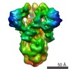 EMDB-9583: 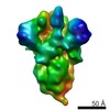 EMDB-9584: 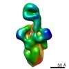 EMDB-9585: 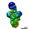 EMDB-9586: 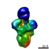 EMDB-9587: EMDB-9588, PDB-6acc: EMDB-9589, PDB-6acd: EMDB-9591, PDB-6acg: EMDB-9593, PDB-6acj: EMDB-9594, PDB-6ack: 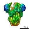 EMDB-9595: 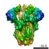 EMDB-9596: 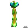 EMDB-9597: 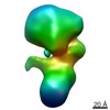 EMDB-9598: |
| Source |
|
 Keywords Keywords | VIRAL PROTEIN / SARS-CoV / spike / glycoprotein / Class I fusion protein / membrane fusion / VIRAL PROTEIN/HYDROLASE / VIRAL PROTEIN-HYDROLASE complex |
 SARS coronavirus
 homo sapiens (human)
 human sars coronavirus