 ムービー
ムービー コントローラー
 構造ビューア
 万見文献について


+検索条件
-Structure paper
| タイトル | Dystrophin's central domain forms a complex filament that becomes disorganized by in-frame deletions. |
|---|---|
| ジャーナル・号・ページ | J Biol Chem, Vol. 293, Issue 18, Page 6637-6646, Year 2018 |
| 掲載日 | 2018年5月4日 |
 著者 著者 | Olivier Delalande / Anne-Elisabeth Molza / Raphael Dos Santos Morais / Angélique Chéron / Émeline Pollet / Céline Raguenes-Nicol / Christophe Tascon / Emmanuel Giudice / Marine Guilbaud / Aurélie Nicolas / Arnaud Bondon / France Leturcq / Nicolas Férey / Marc Baaden / Javier Perez / Pierre Roblin / France Piétri-Rouxel / Jean-François Hubert / Mirjam Czjzek / Elisabeth Le Rumeur /  |
| PubMed 要旨 | Dystrophin, encoded by the gene, is critical for maintaining plasma membrane integrity during muscle contraction events. Mutations in the gene disrupting the reading frame prevent dystrophin ...Dystrophin, encoded by the gene, is critical for maintaining plasma membrane integrity during muscle contraction events. Mutations in the gene disrupting the reading frame prevent dystrophin production and result in severe Duchenne muscular dystrophy (DMD); in-frame internal deletions allow production of partly functional internally deleted dystrophin and result in less severe Becker muscular dystrophy (BMD). Many known BMD deletions occur in dystrophin's central domain, generally considered to be a monotonous rod-shaped domain based on the knowledge of spectrin family proteins. However, the effects caused by these deletions, ranging from asymptomatic to severe BMD, argue against the central domain serving only as a featureless scaffold. We undertook structural studies combining small-angle X-ray scattering and molecular modeling in an effort to uncover the structure of the central domain, as dystrophin has been refractory to characterization. We show that this domain appears to be a tortuous and complex filament that is profoundly disorganized by the most severe BMD deletion (loss of exons 45-47). Despite the preservation of large parts of the binding site for neuronal nitric oxide synthase (nNOS) in this deletion, computational approaches failed to recreate the association of dystrophin with nNOS. This observation is in agreement with a strong decrease of nNOS immunolocalization in muscle biopsies, a parameter related to the severity of BMD phenotypes. The structural description of the whole dystrophin central domain we present here is a first necessary step to improve the design of microdystrophin constructs toward the goal of a successful gene therapy for DMD. |
 リンク リンク | J Biol Chem / PubMed:29535188 / PubMed Central |
| 手法 | SAS (X-ray synchrotron) |
| 構造データ | 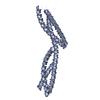 SASDB43: 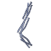 SASDB53:  SASDB63: 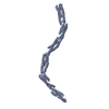 SASDB73:  SASDB83: 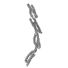 SASDB93: 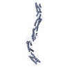 SASDBA3: 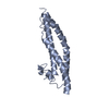 SASDBB3:  SASDBV3: |
| 由来 |
|
 Homo sapiens (ヒト)