 Movie
Movie Controller
 Structure viewers
 About EMN search


-Search query
-Search result
Showing 1 - 50 of 342 items for (author: white & he)

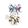
EMDB-44635: 
Inactive mu opioid receptor bound to Nb6, naloxone and NAM
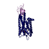
PDB-9bjk:
Inactive mu opioid receptor bound to Nb6, naloxone and NAM
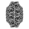
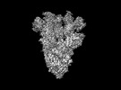
EMDB-16426:
CryoEM structure of the Hendra henipavirus nucleocapsid sauronoid assembly multimer

PDB-8c4h:
CryoEM structure of the Hendra henipavirus nucleocapsid sauronoid assembly multimer

PDB-8cbw:
CryoEM structure of the Hendra henipavirus nucleocapsid sauronoid assembly monomer

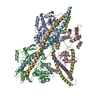
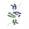
EMDB-42681:
The structure of the native cardiac thin filament troponin core in Ca2+-free state from the upper strand

EMDB-42682:
The structure of the native cardiac thin filament troponin core in Ca2+-free tilted state from the upper strand
EMDB-42683:
The structure of the native cardiac thin filament troponin core in Ca2+-free rotated state from the upper strand

EMDB-42800:
The structure of the native cardiac thin filament troponin core in Ca2+-free state from the lower strand

EMDB-42833:
The structure of the native cardiac thin filament troponin core in Ca2+-free rotated state from the lower strand

EMDB-42835:
The structure of the native cardiac thin filament troponin core in Ca2+-free tilted state from the lower strand

EMDB-42846:
The structure of the native cardiac thin filament troponin core in Ca2+-bound fully activated state from the upper strand

EMDB-42847:
The structure of the native cardiac thin filament troponin core in Ca2+-bound partially activated state from the upper strand

EMDB-42849:
The structure of the native cardiac thin filament troponin core in Ca2+-bound fully activated state 1 from the lower strand

EMDB-42856:
The structure of the native cardiac thin filament troponin core in Ca2+-bound fully activated state 2 from the lower strand

EMDB-42858:
The structure of the native cardiac thin filament troponin core in Ca2+-bound partially activated state from the lower strand

EMDB-42874:
The structure of the native cardiac thin filament troponin core in Ca2+-free state from the upper strand activated by the C1-domain of cardiac myosin binding protein C

PDB-8uww:
The structure of the native cardiac thin filament troponin core in Ca2+-free state from the upper strand

PDB-8uwx:
The structure of the native cardiac thin filament troponin core in Ca2+-free tilted state from the upper strand
PDB-8uwy:
The structure of the native cardiac thin filament troponin core in Ca2+-free rotated state from the upper strand

PDB-8uyd:
The structure of the native cardiac thin filament troponin core in Ca2+-free state from the lower strand

PDB-8uz5:
The structure of the native cardiac thin filament troponin core in Ca2+-free rotated state from the lower strand

PDB-8uz6:
The structure of the native cardiac thin filament troponin core in Ca2+-free tilted state from the lower strand

PDB-8uzx:
The structure of the native cardiac thin filament troponin core in Ca2+-bound fully activated state from the upper strand

PDB-8uzy:
The structure of the native cardiac thin filament troponin core in Ca2+-bound partially activated state from the upper strand

PDB-8v01:
The structure of the native cardiac thin filament troponin core in Ca2+-bound fully activated state 1 from the lower strand

PDB-8v0i:
The structure of the native cardiac thin filament troponin core in Ca2+-bound fully activated state 2 from the lower strand

PDB-8v0k:
The structure of the native cardiac thin filament troponin core in Ca2+-bound partially activated state from the lower strand

PDB-8v0y:
The structure of the native cardiac thin filament troponin core in Ca2+-free state from the upper strand activated by the C1-domain of cardiac myosin binding protein C
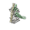
EMDB-28198:
Cryo-EM map of SARS-CoV-2 Omicron BA.2 spike in complex with LLNL-199

EMDB-28199:
Cryo-EM map of SARS-CoV-2 Omicron BA.2 spike in complex with 2130-1-0114-112
PDB-8ekd:
Cryo-EM map of SARS-CoV-2 Omicron BA.2 spike in complex with 2130-1-0114-112
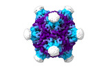
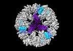
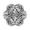
EMDB-19024:
Structure of the PNMA2 capsid
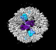
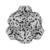
EMDB-19025:
Structure of the five-fold capsomer of the PNMA2 capsid
EMDB-19026:
Structure of the three-fold capsomer of the PNMA2 capsid
EMDB-19027:
Structure of the two-fold capsomer of the PNMA2 capsid
PDB-8rb3:
Structure of the PNMA2 capsid

PDB-8rb4:
Structure of the five-fold capsomer of the PNMA2 capsid
PDB-8rb5:
Structure of the three-fold capsomer of the PNMA2 capsid
PDB-8rb7:
Structure of the two-fold capsomer of the PNMA2 capsid
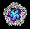
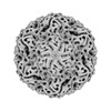
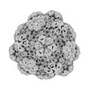
EMDB-43013:
Myxococcus xanthus HEnc-K417N(A) protein shell with icosahedral T=1 symmetry

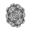
EMDB-43016:
Myxococcus xanthus HEnc-K417N(A) protein shell with tetrahedral symmetry (12 pentamers, 4 hexamers)
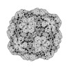
EMDB-43037:
Myxococcus xanthus HEnc-K417N(A) protein shell with D3 symmetry (12 pentamers, 3 hexamers)
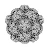
EMDB-43038:
Myxococcus xanthus HEnc-K417N(A) protein shell with D6 symmetry (12 pentamers, 8 hexamers)

EMDB-43039:
Myxococcus xanthus HEnc-K417N(A) protein shell with C2 symmetry (12 pentamers, 9 hexamers)
EMDB-43040:
Myxococcus xanthus HEnc-K417N(A) protein shell with D3 symmetry (12 pentamers, 11 hexamers)
EMDB-43041:
Myxococcus xanthus HEnc-K417N(A) protein shell with D2 symmetry (12 pentamers, 12 hexamers)
EMDB-43042:
Myxococcus xanthus HEnc-K417N(A) protein shell with D2 symmetry (12 pentamers, 14 hexamers)
EMDB-43043:
Myxococcus xanthus HEnc-K417N(A) protein shell with D5 symmetry (12 pentamers, 15 hexamers)
EMDB-43113:
Myxococcus xanthus EncA WT protein shell with icosahedral symmetry T=3
Pages:
 wwPDB to switch to version 3 of the EMDB data model
