 Movie
Movie Controller

[English] 日本語
 Yorodumi
- PDB-8c4h: CryoEM structure of the Hendra henipavirus nucleocapsid sauronoid... -
+ Open data

- Basic information
| Entry | Database: PDB / ID: 8c4h | ||||||
|---|---|---|---|---|---|---|---|
| Title | CryoEM structure of the Hendra henipavirus nucleocapsid sauronoid assembly multimer | ||||||
 Components Components |
| ||||||
 Keywords Keywords | VIRAL PROTEIN / Nucleoprotein / RNA-binding protein / Sauronoid | ||||||
| Function / homology |  Function and homology information Function and homology informationhelical viral capsid / viral nucleocapsid / host cell cytoplasm / ribonucleoprotein complex / structural molecule activity / RNA binding Similarity search - Function | ||||||
| Biological species |  Hendra henipavirus Hendra henipavirus | ||||||
| Method | ELECTRON MICROSCOPY / single particle reconstruction / cryo EM / Resolution: 3.485 Å | ||||||
 Authors Authors | Passchier, T.C. / Maskell, D.P. / Edwards, T.A. / Barr, J.N. | ||||||
| Funding support | European Union, 1items
| ||||||
 Citation Citation | Journal: Sci Rep / Year: 2024 Title: The cryoEM structure of the Hendra henipavirus nucleoprotein reveals insights into paramyxoviral nucleocapsid architectures. Authors: Tim C Passchier / Joshua B R White / Daniel P Maskell / Matthew J Byrne / Neil A Ranson / Thomas A Edwards / John N Barr /   Abstract: We report the first cryoEM structure of the Hendra henipavirus nucleoprotein in complex with RNA, at 3.5 Å resolution, derived from single particle analysis of a double homotetradecameric RNA-bound ...We report the first cryoEM structure of the Hendra henipavirus nucleoprotein in complex with RNA, at 3.5 Å resolution, derived from single particle analysis of a double homotetradecameric RNA-bound N protein ring assembly exhibiting D14 symmetry. The structure of the HeV N protein adopts the common bi-lobed paramyxoviral N protein fold; the N-terminal and C-terminal globular domains are bisected by an RNA binding cleft containing six RNA nucleotides and are flanked by the N-terminal and C-terminal arms, respectively. In common with other paramyxoviral nucleocapsids, the lateral interface between adjacent N and N protomers involves electrostatic and hydrophobic interactions mediated primarily through the N-terminal arm and globular domains with minor contribution from the C-terminal arm. However, the HeV N multimeric assembly uniquely identifies an additional protomer-protomer contact between the N N-terminus and N C-terminal arm linker. The model presented here broadens the understanding of RNA-bound paramyxoviral nucleocapsid architectures and provides a platform for further insight into the molecular biology of HeV, as well as the development of antiviral interventions. | ||||||
| History |
|
- Structure visualization
| Structure viewer | Molecule: MolmilJmol/JSmol |
|---|
- Downloads & links
-Download
| PDBx/mmCIF format | 8c4h.cif.gz | 2 MB | Display | PDBx/mmCIF format |
|---|---|---|---|---|
| PDB format | pdb8c4h.ent.gz | 1.7 MB | Display | PDB format |
| PDBx/mmJSON format | 8c4h.json.gz | Tree view | PDBx/mmJSON format | |
| Others |  Other downloads Other downloads |
-Validation report
| Arichive directory | https://data.pdbj.org/pub/pdb/validation_reports/c4/8c4hftp://data.pdbj.org/pub/pdb/validation_reports/c4/8c4h | HTTPS FTP |
|---|
-Related structure data
| Related structure data | 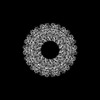 16426MC  8cbwC M: map data used to model this data C: citing same article ( |
|---|---|
| Similar structure data |
-Links
 PDBj
PDBj


- Assembly
| Deposited unit | 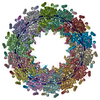
|
|---|---|
| 1 |
|
-Components
| #1: Protein | Mass: 58541.320 Da / Num. of mol.: 28 Source method: isolated from a genetically manipulated source Source: (gene. exp.) Hendra henipavirus / Plasmid: pET28a / Production host: #2: RNA chain | Mass: 25672.984 Da / Num. of mol.: 2 Source method: isolated from a genetically manipulated source Source: (gene. exp.) |
|---|
-Experimental details
-Experiment
| Experiment | Method: ELECTRON MICROSCOPY |
|---|---|
| EM experiment | Aggregation state: PARTICLE / 3D reconstruction method: single particle reconstruction |
- Sample preparation
| Component | Name: Sauronoid assembly of Hendra henipavirus nucleoprotein Type: COMPLEX Details: Recombinantly expressed Hendra henipavirus nucleoprotein forming sauronoid complexes. Entity ID: all / Source: RECOMBINANT |
|---|---|
| Molecular weight | Value: 1.691779 MDa / Experimental value: NO |
| Source (natural) | Organism: Hendra henipavirus |
| Source (recombinant) | Organism: |
| Details of virus | Isolate: SPECIES / Type: VIRION |
| Buffer solution | pH: 7.5 |
| Specimen | Embedding applied: NO / Shadowing applied: NO / Staining applied: NO / Vitrification applied: YES |
| Specimen support | Grid material: COPPER / Grid type: PELCO Ultrathin Carbon with Lacey Carbon |
| Vitrification | Instrument: FEI VITROBOT MARK IV / Cryogen name: ETHANE / Humidity: 95 % / Chamber temperature: 281 K |
- Electron microscopy imaging
| Experimental equipment |  Model: Titan Krios / Image courtesy: FEI Company |
|---|---|
| Microscopy | Model: FEI TITAN KRIOS |
| Electron gun | Electron source:  FIELD EMISSION GUN / Accelerating voltage: 300 kV / Illumination mode: FLOOD BEAM FIELD EMISSION GUN / Accelerating voltage: 300 kV / Illumination mode: FLOOD BEAM |
| Electron lens | Mode: BRIGHT FIELD / Nominal magnification: 75000 X / Nominal defocus max: 2800 nm / Nominal defocus min: 700 nm |
| Specimen holder | Cryogen: NITROGEN / Specimen holder model: FEI TITAN KRIOS AUTOGRID HOLDER |
| Image recording | Average exposure time: 1.5 sec. / Electron dose: 59.7 e/Å2 / Detector mode: INTEGRATING / Film or detector model: FEI FALCON III (4k x 4k) / Num. of grids imaged: 1 / Num. of real images: 3548 |
- Processing
| EM software |
| ||||||||||||||||||||||||||||||||
|---|---|---|---|---|---|---|---|---|---|---|---|---|---|---|---|---|---|---|---|---|---|---|---|---|---|---|---|---|---|---|---|---|---|
| CTF correction | Type: PHASE FLIPPING AND AMPLITUDE CORRECTION | ||||||||||||||||||||||||||||||||
| Symmetry | Point symmetry: D14 (2x14 fold dihedral) | ||||||||||||||||||||||||||||||||
| 3D reconstruction | Resolution: 3.485 Å / Resolution method: FSC 0.143 CUT-OFF / Num. of particles: 5683 / Num. of class averages: 1 / Symmetry type: POINT | ||||||||||||||||||||||||||||||||
| Refine LS restraints |
|
