 ムービー
ムービー コントローラー
 構造ビューア
 EMN検索について


-検索条件
-検索結果
検索 (著者・登録者: weninger & g)の結果全44件を表示しています

EMDBエントリ 画像なし
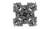
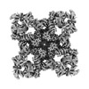
EMDB-43296: 
Constituent EM map: Focused refinement S100A1 of mouse RyR1 in complex with S100A1 (EGTA-only dataset)
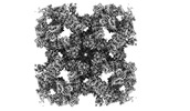
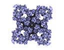
EMDB-43304:
Structure of mouse RyR1 in complex with S100A1 (high-Ca2+/CFF/ATP dataset)

PDB-8vk4:
Structure of mouse RyR1 in complex with S100A1 (high-Ca2+/CFF/ATP dataset)
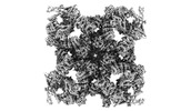
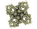
EMDB-43299:
Structure of mouse RyR1 in complex with S100A1 (EGTA-only dataset)

PDB-8vk3:
Structure of mouse RyR1 in complex with S100A1 (EGTA-only dataset)

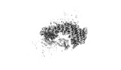
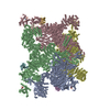
EMDB-43283:
Structure of mouse RyR1 (EGTA-only dataset)

EMDB-43284:
Structure of mouse RyR1 (high-Ca2+/CFF/ATP dataset)
EMDB-43295:
Raw consensus map of mouse RyR1 in complex with S100A1 (EGTA-only dataset)
EMDB-43297:
Constituent EM map: Focused refinement BSol+S100A1 of mouse RyR1 in complex with S100A1 (EGTA-only dataset)
EMDB-43298:
Constituent EM map: Focused refinement JSol+CSol+S100A1 of mouse RyR1 in complex with S100A1 (EGTA-only dataset)
EMDB-43300:
Raw consensus map of mouse RyR1 in complex with S100A1 (high-Ca2+/CFF/ATP dataset)
EMDB-43301:
Constituent EM map: Focused refinement JSol+CSol+S100A1 of mouse RyR1 in complex with S100A1 (high-Ca2+/CFF/ATP dataset)
EMDB-43302:
Constituent EM map: Focused refinement BSol+S100A1 of mouse RyR1 in complex with S100A1 (high-Ca2+/CFF/ATP dataset)
EMDB-43303:
Constituent EM map: Focused refinement S100A1 of mouse RyR1 in complex with S100A1 (high-Ca2+/CFF/ATP dataset)
EMDB-43307:
Raw consensus map of mouse RyR1 (EGTA-only dataset)
EMDB-43308:
Constituent EM map: Focused refinement NTD+SPRY+Calstabin-1 of mouse RyR1 (EGTA-only dataset)
EMDB-43309:
Constituent EM map: Focused refinement JSol+CSol of mouse RyR1 (EGTA-only dataset)
EMDB-43310:
Constituent EM map: Focused refinement BSol of mouse RyR1 (EGTA-only dataset)
EMDB-43311:
Constituent EM map: Focused refinement TaF+TM+CTD of mouse RyR1 (EGTA-only dataset)
EMDB-43312:
Constituent EM map: Focused refinement Ry1&2 of mouse RyR1 (EGTA-only dataset)
EMDB-43313:
Constituent EM map: Focused refinement Ry3&4 of mouse RyR1 (EGTA-only dataset)

PDB-8vjj:
Structure of mouse RyR1 (EGTA-only dataset)

PDB-8vjk:
Structure of mouse RyR1 (high-Ca2+/CFF/ATP dataset)
EMDB-26139:
Structure of human RyR2 in the closed state.

EMDB-26405:
Structure of PKA phosphorylated human RyR2 in the closed state

EMDB-26407:
Structure of PKA phosphorylated human RyR2 in the open state

EMDB-26408:
Structure of PKA phosphorylated human RyR2 in the closed state in the presence of Calmodulin
EMDB-26409:
Structure of PKA phosphorylated human RyR2-R2474S in the closed state

EMDB-26410:
Structure of PKA phosphorylated human RyR2-R2474S in the open state

EMDB-26412:
Structure of PKA phosphorylated human RyR2-R2474S in the closed state in the presence of ARM210

EMDB-26413:
Structure of PKA phosphorylated human RyR2-R2474S in the closed state in the presence of Calmodulin

EMDB-26414:
Structure of PKA phosphorylated human RyR2-R2474S in the open state in the presence of Calmodulin

EMDB-26415:
Structure of dephosphorylated human RyR2 in the closed state
EMDB-26416:
Structure of dephosphorylated human RyR2 in the open state

PDB-7u9q:
Structure of PKA phosphorylated human RyR2 in the closed state

PDB-7u9r:
Structure of PKA phosphorylated human RyR2 in the open state

PDB-7u9t:
Structure of PKA phosphorylated human RyR2 in the closed state in the presence of Calmodulin

PDB-7u9x:
Structure of PKA phosphorylated human RyR2-R2474S in the closed state

PDB-7u9z:
Structure of PKA phosphorylated human RyR2-R2474S in the open state

PDB-7ua1:
Structure of PKA phosphorylated human RyR2-R2474S in the closed state in the presence of ARM210

PDB-7ua3:
Structure of PKA phosphorylated human RyR2-R2474S in the closed state in the presence of Calmodulin

PDB-7ua4:
Structure of PKA phosphorylated human RyR2-R2474S in the open state in the presence of Calmodulin
PDB-7ua5:
Structure of dephosphorylated human RyR2 in the closed state

PDB-7ua9:
Structure of dephosphorylated human RyR2 in the open state
 wwPDBはEMDBデータモデルのバージョン3へ移行します
