 ムービー
ムービー コントローラー
 構造ビューア
 EMN検索について


-検索条件
-検索結果
検索 (著者・登録者: ventura & s)の結果全26件を表示しています

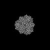
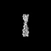
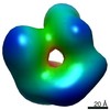
EMDB-16466: 
The structural architecture of alpha-synuclein oligomer

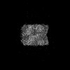
EMDB-16528:
3D reconstruction of alpha-synuclein oligomer-PSMa3 complex
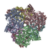
EMDB-16521:
Cryo-EM structure of the Cibeles-Demetra 3:3 heterocomplex from Galleria mellonella saliva
EMDB-16524:
Cryo-EM structure of the Ceres homohexamer from Galleria mellonella saliva

EMDB-16531:
Cryo-EM structure of the Cora homohexamer from Galleria mellonella saliva
PDB-8ca9:
Cryo-EM structure of the Cibeles-Demetra 3:3 heterocomplex from Galleria mellonella saliva

PDB-8cad:
Cryo-EM structure of the Ceres homohexamer from Galleria mellonella saliva

PDB-8can:
Cryo-EM structure of the Cora homohexamer from Galleria mellonella saliva
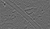
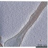
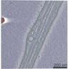
EMDB-18475:
Cryo-electron tomogram of an induced S2 cell protrusion. The cell was treated with 2uM thapsigargin (5h) and with 2.5uM Cytochalasin D (2h).
EMDB-16877:
Subtomogram averaging structure of cofilactin filament inside microtubule lumen of Drosophila S2 cell protrusion.

PDB-8oh4:
Subtomogram averaging structure of cofilactin filament inside microtubule lumen of Drosophila S2 cell protrusion.
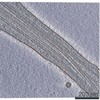
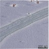
EMDB-16685:
Tomogram of an induced protrusion of a Drosophila S2 cell
EMDB-16693:
Tomogram of an induced protrusion of a Drosophila S2 cell
EMDB-16695:
Tomogram of an induced protrusion of a Drosophila S2 alpha-tubulin acetyltransferase knock-out (dTAT KO) cell with a filament inside the microtubule lumen
EMDB-16720:
Tomogram of an induced protrusion of a Drosophila S2 cell with filaments inside the microtubule lumen.
EMDB-16800:
Tomogram of an induced protrusion of a cofilin knock-down Drosophila S2 cell with filaments inside the microtubule lumen
EMDB-16811:
Tomogram of an induced protrusion of a cofilin knock-down Drosophila S2 cell with a filament inside the microtubule lumen.

EMDB-14738:
Cryo-EM structure of hnRNPDL amyloid fibrils
PDB-7zir:
Cryo-EM structure of hnRNPDL amyloid fibrils

EMDB-12639:
In situ subtomogram average of 13 protofilament microtubule from Mus musculus DRG axons
EMDB-12640:
In situ subtomogram average of microtubule inner protein from Mus musculus DRG axons
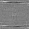
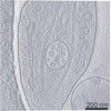
EMDB-13598:
Tomogram of a mouse dorsal root ganglion axon (dataset 2, TS_43).
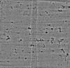
EMDB-13599:
Tomogram of mouse dorsal root ganglion axon (dataset 2, TS_41).
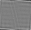
EMDB-13600:
Tomogram of mouse dorsal root ganglion axon (dataset 1, TS_14).
EMDB-13601:
Tomogram of mouse dorsal root ganglion axon (dataset 1, TS_20).
EMDB-13602:
Tomogram of mouse dorsal root ganglion axon (dataset 1, TS_29).
 wwPDBはEMDBデータモデルのバージョン3へ移行します
