 Movie
Movie Controller
 Structure viewers
 About EMN search


-Search query
-Search result
Showing 1 - 50 of 51 items for (author: seitz & s)

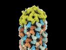
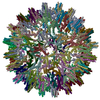
EMDB-16076: 
Helical shell of CCMV capsid protein on DNA origami 6HB-2k

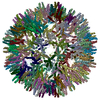
EMDB-16077:
Inner helical shell of CCMV capsid protein on DNA origami 6HB-10k

EMDB-16078:
Outer helical shell of CCMV capsid protein on DNA origami 6HB-10k
EMDB-16079:
Helical shell cap of CCMV capsid protein on DNA origami 6HB-2k

EMDB-16080:
Helical shell of CCMV capsid protein on DNA origami 24HB-2.5k
PDB-8bi4:
Helical shell of CCMV capsid protein on DNA origami 6HB-2k
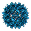
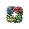
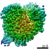
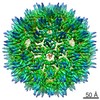
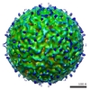
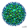
EMDB-15295:
African cichlid nackednavirus capsid at pH 7.5
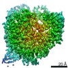
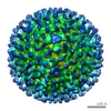
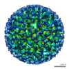
EMDB-16371:
African cichlid nackednavirus capsid at pH 5.5
PDB-8aac:
African cichlid nackednavirus capsid at pH 7.5
PDB-8c0o:
African cichlid nackednavirus capsid at pH 5.5

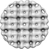
EMDB-25183:
P. chlororaphis 70S ribosome in situ subtomogram average

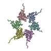
EMDB-25220:
In situ subtomogram average of the 201phi2-1 phage nucleus major shell protein, chimallin (concave class)
EMDB-25221:
In situ consensus subtomogram average of the 201phi2-1 chimallin

EMDB-25222:
In situ subtomogram average of 201phi2-1 phage nucleus major shell protein, chimallin (intermediate/flat class)

EMDB-25223:
In situ subtomogram average of the 201phi2-1 phage nucleus major shell protein, chimallin (convex class)

EMDB-25229:
In situ subtomogram average of the Goslar major phage nucleus shell protein, chimallin (consensus class)

EMDB-25262:
In situ subtomogram average of Goslar phage nucleus major shell protein, chimallin (concave class)

EMDB-25358:
In situ subtomogram average of the major Goslar phage nucleus shell protein, chimallin (convex class)

EMDB-25359:
In situ subtomogram average of the APEC2248 70S ribosome

EMDB-25360:
In situ subtomogram average of the APEC2248 50S ribosome

EMDB-25390:
201Phi2-1 Chimallin Cubic (O, 24mer) assembly
EMDB-25391:
201phi2-1 Chimallin localized tetramer reconstruction
EMDB-25392:
201phi2-1 Chimallin C1 localized reconstruction

EMDB-25393:
201phi2-1 chimallin rectangular (D4,40mer) assembly

EMDB-25394:
Goslar chimallin cubic (O, 24mer) assembly

EMDB-25395:
Goslar chimallin C4 tetramer localized reconstruction

EMDB-25396:
Goslar chimallin C1 localized reconstruction
PDB-7sqq:
201Phi2-1 Chimallin Cubic (O, 24mer) assembly
PDB-7sqr:
201phi2-1 Chimallin localized tetramer reconstruction
PDB-7sqs:
201phi2-1 Chimallin C1 localized reconstruction
PDB-7sqt:
Goslar chimallin cubic (O, 24mer) assembly
PDB-7squ:
Goslar chimallin C4 tetramer localized reconstruction
PDB-7sqv:
Goslar chimallin C1 localized reconstruction
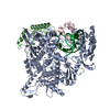
EMDB-11993:
Structure of elongating SARS-CoV-2 RNA-dependent RNA polymerase with Remdesivir at position -3 (structure 1)
EMDB-11994:
Structure of elongating SARS-CoV-2 RNA-dependent RNA polymerase with Remdesivir at position -4 (structure 2)
EMDB-11995:
Structure of elongating SARS-CoV-2 RNA-dependent RNA polymerase with AMP at position -4 (structure 3)
PDB-7b3b:
Structure of elongating SARS-CoV-2 RNA-dependent RNA polymerase with Remdesivir at position -3 (structure 1)
PDB-7b3c:
Structure of elongating SARS-CoV-2 RNA-dependent RNA polymerase with Remdesivir at position -4 (structure 2)
PDB-7b3d:
Structure of elongating SARS-CoV-2 RNA-dependent RNA polymerase with AMP at position -4 (structure 3)
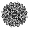
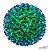
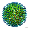
EMDB-3822:
Structure of the full-length African cichlid nackednavirus icosahedral capsid

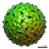
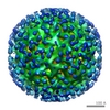
EMDB-3823:
Structure of the truncated African cichlid nackednavirus icosahedral capsid
EMDB-1399:
Cryo-electron microscopy of hepatitis B virions reveals variability in envelope capsid interactions.

EMDB-1400:
Cryo-electron microscopy of hepatitis B virions reveals variability in envelope capsid interactions.
EMDB-1401:
Cryo-electron microscopy of hepatitis B virions reveals variability in envelope capsid interactions.
EMDB-1402:
Cryo-electron microscopy of hepatitis B virions reveals variability in envelope capsid interactions.
EMDB-1403:
Cryo-electron microscopy of hepatitis B virions reveals variability in envelope capsid interactions.
EMDB-1404:
Cryo-electron microscopy of hepatitis B virions reveals variability in envelope capsid interactions.
EMDB-1405:
Cryo-electron microscopy of hepatitis B virions reveals variability in envelope capsid interactions.
EMDB-1406:
Cryo-electron microscopy of hepatitis B virions reveals variability in envelope capsid interactions.
EMDB-1407:
Cryo-electron microscopy of hepatitis B virions reveals variability in envelope capsid interactions.
Pages:
 wwPDB to switch to version 3 of the EMDB data model
