 Movie
Movie Controller
 Structure viewers
 About EMN search


-Search query
-Search result
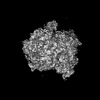
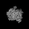
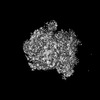
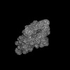
Showing 1 - 50 of 85 items for (author: naismith & jh)

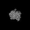
EMDB Unreleased entry
EMDB-54064: 
Structure of the O-oligosaccharyl transferase PglL from Neisseria meningitidis in complex with a nanobody
Method: single particle / : Harrison PJ, Naismith JH, Clare DK, Quigley A

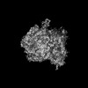
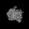
EMDB-54649:
Structure of E. coli WbaP in complex with a megabody
Method: single particle / : Weckener M, Le Bas A, Ward PN, Naismith JH

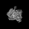
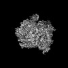
EMDB-54664:
Structure of E. coli WbaP in complex with a megabody in presence of UDP-Gal
Method: single particle / : Weckener M, Le Bas A, Ward PN, Naismith JH

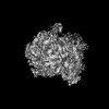
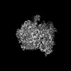
EMDB-58070:
Structure of E. coli WbaP in complex with a megabody
Method: single particle / : Weckener M, Le Bas A, Ward PN, Naismith JH

EMDB-58108:
Structure of E. coli WbaP in complex with a megabody in the presence of UDP-Gal processed in C2 symmetry
Method: single particle / : Weckener M, Le Bas A, Ward PN, Harrison PJ, Naismith JH

EMDB-51917:
Streptavidin map obtained from graphene oxide modified self-wicking grids
Method: single particle / : Weckener M, Darrow MC, Clare DK, Naismith JH

EMDB-51440:
3D reconstruction of beta-galactosidase obtained from self-wicking grids
Method: single particle / : Weckener M, Clare DK, Darrow MC, Naismith JH

EMDB-51415:
3D reconstruction of beta-galactosidase obtained from graphene oxide modified self-wicking grids
Method: single particle / : Weckener M, Clare DK, Darrow MC, Naismith JH

EMDB-50042:
Wzc-K540M-2YE MgADP C1
Method: single particle / : Liu JW, Yang Y, Naismith JH

EMDB-50043:
Wzc-K540M-2YE MgADP C8
Method: single particle / : Liu JW, Yang Y, Naismith JH

EMDB-50044:
Wzc-K540M-3YE MgADP C1
Method: single particle / : Liu JW, Yang Y, Naismith JH

EMDB-50045:
Wzc-K540M-3YE MgADP C8
Method: single particle / : Liu JW, Yang Y, Naismith JH

EMDB-50046:
Wzc-K540M-3YE-N711Y MgADP C1
Method: single particle / : Liu JW, Yang Y, Naismith JH

EMDB-50047:
Wzc-K540M-3YE-N711Y MgADP C8
Method: single particle / : Liu JW, Yang Y, Naismith JH
EMDB-50675:
Escherichia coli 70S ribosome in situ structure
Method: subtomogram averaging / : Watson H, Berger C, Grange M
EMDB-50676:
E. coli 70S ribosome in situ structure for xenon damage layer determination: 5 - 10 nm
Method: subtomogram averaging / : Watson H, Berger C, Grange M
EMDB-50678:
E. coli 70S ribosome in situ structure for xenon damage layer determination: 10 - 15 nm
Method: subtomogram averaging / : Watson H, Berger C, Grange M
EMDB-50679:
E. coli 70S ribosome in situ structure for xenon damage layer determination: 15 - 20 nm
Method: subtomogram averaging / : Watson H, Berger C, Grange M

EMDB-50680:
E. coli 70S ribosome in situ structure for xenon damage layer determination: 20 - 25 nm
Method: subtomogram averaging / : Watson H, Berger C, Grange M

EMDB-50681:
E. coli 70S ribosome in situ structure for xenon damage layer determination: 25 - 30 nm
Method: subtomogram averaging / : Watson H, Berger C, Grange M
EMDB-50682:
E. coli 70S ribosome in situ structure for xenon damage layer determination: 30 - 35 nm
Method: subtomogram averaging / : Watson H, Berger C, Grange M
EMDB-50683:
E. coli 70S ribosome in situ structure for xenon damage layer determination: 35 - 40 nm
Method: subtomogram averaging / : Watson H, Berger C, Grange M
EMDB-50684:
E. coli 70S ribosome in situ structure for xenon damage layer determination: 40 - 45 nm
Method: subtomogram averaging / : Watson H, Berger C, Grange M
EMDB-50685:
E. coli 70S ribosome in situ structure for xenon damage layer determination: 45 - 50 nm
Method: subtomogram averaging / : Watson H, Berger C, Grange M
EMDB-50686:
E. coli 70S ribosome in situ structure for xenon damage layer determination: 50 - 55 nm
Method: subtomogram averaging / : Watson H, Berger C, Grange M

EMDB-50687:
E. coli 70S ribosome in situ structure for xenon damage layer determination: 55 - 60 nm
Method: subtomogram averaging / : Watson H, Berger C, Grange M
EMDB-50688:
E. coli 70S ribosome in situ structure for xenon damage layer determination: >10nm matched control for 5 - 10 nm
Method: subtomogram averaging / : Watson H, Berger C, Grange M

EMDB-50689:
E. coli 70S ribosome in situ structure for xenon damage layer determination: >15 nm matched control for 10 - 15 nm
Method: subtomogram averaging / : Watson H, Berger C, Grange M

EMDB-50690:
E. coli 70S ribosome in situ structure for xenon damage layer determination: >20 nm matched control for 15 - 20 nm
Method: subtomogram averaging / : Watson H, Berger C, Grange M

EMDB-50691:
E. coli 70S ribosome in situ structure for xenon damage layer determination: >25 nm matched control for 20 - 25 nm
Method: subtomogram averaging / : Watson H, Berger C, Grange M

EMDB-50692:
E. coli 70S ribosome in situ structure for xenon damage layer determination: >30 nm matched control for 25 - 30 nm
Method: subtomogram averaging / : Watson H, Berger C, Grange M

EMDB-50693:
E. coli 70S ribosome in situ structure for xenon damage layer determination: >35 nm matched control for 30 - 35 nm
Method: subtomogram averaging / : Watson H, Berger C, Grange M

EMDB-50694:
E. coli 70S ribosome in situ structure for xenon damage layer determination: >40 nm matched control for 35 - 40 nm
Method: subtomogram averaging / : Watson H, Berger C, Grange M

EMDB-50695:
E. coli 70S ribosome in situ structure for xenon damage layer determination: >45 nm matched control for 40 - 45 nm
Method: subtomogram averaging / : Watson H, Berger C, Grange M

EMDB-50696:
E. coli 70S ribosome in situ structure for xenon damage layer determination: >50 nm matched control for 45 - 50 nm
Method: subtomogram averaging / : Watson H, Berger C, Grange M

EMDB-50697:
E. coli 70S ribosome in situ structure for xenon damage layer determination: >55 nm matched control for 50 - 55 nm
Method: subtomogram averaging / : Watson H, Berger C, Grange M

EMDB-50698:
E. coli 70S ribosome in situ structure for xenon damage layer determination: >60 nm matched control for 55 - 60 nm
Method: subtomogram averaging / : Watson H, Berger C, Grange M

EMDB-50699:
E. coli 70S ribosome in situ structure for lamellae backside damage analysis: 0 - 1 micron from lamellae backside
Method: subtomogram averaging / : Watson H, Berger C, Grange M

EMDB-50700:
E. coli 70S ribosome in situ structure for lamellae backside damage analysis: 1 - 2 microns from lamellae backside
Method: subtomogram averaging / : Watson H, Berger C, Grange M

EMDB-50701:
E. coli 70S ribosome in situ structure for lamellae backside damage analysis: 2 - 3 microns from lamellae backside
Method: subtomogram averaging / : Watson H, Berger C, Grange M

EMDB-50702:
E. coli 70S ribosome in situ structure for lamellae backside damage analysis: 3 - 4 microns from lamellae backside
Method: subtomogram averaging / : Watson H, Berger C, Grange M

EMDB-50703:
E. coli 70S ribosome in situ structure for lamellae backside damage analysis: 4 - 5 microns from lamellae backside
Method: subtomogram averaging / : Watson H, Berger C, Grange M

EMDB-50704:
E. coli 70S ribosome in situ structure for xenon damage layer determination: 10,000 particles (no depth constraint)
Method: subtomogram averaging / : Watson H, Berger C, Grange M

EMDB-50705:
E. coli 70S ribosome in situ structure for xenon damage layer determination: 10,000 particles (>45 nm)
Method: subtomogram averaging / : Watson H, Berger C, Grange M

EMDB-50706:
E. coli 70S ribosome in situ structure for xenon damage layer determination: 10,000 particles (<=45 nm)
Method: subtomogram averaging / : Watson H, Berger C, Grange M

EMDB-52177:
E. coli 70S ribosome in situ structure for xenon damage layer determination: 10,000 particles (no depth constraint)
Method: subtomogram averaging / : Watson H, Berger C, Grange M

EMDB-52178:
E. coli 70S ribosome in situ structure for xenon damage layer determination: 10,000 particles (<30 nm)
Method: subtomogram averaging / : Watson H, Berger C, Grange M

EMDB-52179:
E. coli 70S ribosome in situ structure for xenon damage layer determination: 10,000 particles (>30 nm)
Method: subtomogram averaging / : Watson H, Berger C, Grange M
EMDB-17295:
Stabilised BA.1 SARS-CoV-2 spike with H6 nanobodies in '3 up' RBD conformation
Method: single particle / : Weckener M, Naismith JH, Owens RJ

EMDB-17296:
Stabilised BA.1 SARS-CoV-2 spike with H6 nanobodies in '2 up 1 down' RBD conformation
Method: single particle / : Weckener M, Naismith JH, Owens RJ
Pages:
 wwPDB to switch to version 3 of the EMDB data model
