 Movie
Movie Controller
 Structure viewers
 About EMN search


-Search query
-Search result
Showing all 43 items for (author: moser & f)

EMDB-76979: 
Cryo-ET of mitochondrial membrane in direct interaction with alpha-synuclein exhibiting membrane morphological distortion
Method: electron tomography / : Jaber N, Dai W

EMDB-76980:
Supplemental: irregularly shaped mitochondria interacting with alpha-synuclein
Method: electron tomography / : Jaber N, Dai W

EMDB-76981:
Supplemental: alpha-synuclein oligomers on the surface of a mitochondrial membrane
Method: electron tomography / : Jaber N, Dai W

EMDB-76983:
Supplemental: mitochondria not associated with alpha-synuclein
Method: electron tomography / : Jaber N, Dai W

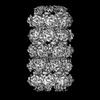
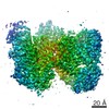
EMDB-53046:
Mouse otoferlin (216-1931) in complex with an MSP2N2 lipid nanodisc (30 mol% DOPS, 10 mol% PI(4,5)P2)
Method: single particle / : Cretu C, Moser T

PDB-9qe2:
Mouse otoferlin (216-1931) in complex with an MSP2N2 lipid nanodisc (30 mol% DOPS, 10 mol% PI(4,5)P2)
Method: single particle / : Cretu C, Moser T

EMDB-54802:
Mouse otoferlin (216-1931) in the lipid-free, Ca2+-bound state, "open" conformation (class 2)
Method: single particle / : Cretu C, Moser T

EMDB-54805:
Mouse otoferlin (216-1931) in complex with a lipid nanodisc (comprising 25% PS and 5% PIP2)
Method: single particle / : Cretu C, Moser T

EMDB-54809:
Mouse otoferlin (216-1931) in the lipid-free Ca2+-bound state, "open" conformation (class 1)
Method: single particle / : Cretu C, Moser T

EMDB-54827:
Mouse otoferlin (residues 216-1931) in the lipid-bound state (merged datasets)
Method: single particle / : Cretu C, Moser T

EMDB-54883:
Mouse otoferlin (216-1931) in the lipid-free, Ca2+-free state ("loose" conformation)
Method: single particle / : Cretu C, Moser T

EMDB-54923:
Mouse otoferlin (216-1931) in the lipid-free Ca2+-bound state, "closed-like" conformation
Method: single particle / : Cretu C, Moser T

PDB-9se5:
Mouse otoferlin (216-1931) in the lipid-free, Ca2+-bound state, "open" conformation (class 2)
Method: single particle / : Cretu C, Moser T

PDB-9sea:
Mouse otoferlin (216-1931) in complex with a lipid nanodisc (comprising 25% PS and 5% PIP2)
Method: single particle / : Cretu C, Moser T

PDB-9seg:
Mouse otoferlin (216-1931) in the lipid-free Ca2+-bound state, "open" conformation (class 1)
Method: single particle / : Cretu C, Moser T

PDB-9sfl:
Mouse otoferlin (residues 216-1931) in the lipid-bound state (merged datasets)
Method: single particle / : Cretu C, Moser T

PDB-9sh0:
Mouse otoferlin (216-1931) in the lipid-free, Ca2+-free state ("loose" conformation)
Method: single particle / : Cretu C, Moser T

PDB-9si1:
Mouse otoferlin (216-1931) in the lipid-free Ca2+-bound state, "closed-like" conformation
Method: single particle / : Cretu C, Moser T

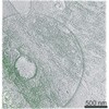
EMDB-53557:
Architecture of the Drosophila nephrocyte slit diaphragm revealed by cryo-electron tomography
Method: subtomogram averaging / : Moser D, Scheffer MP, Frangakis AF

EMDB-18795:
Cryo-EM structure of the microbial rhodopsin CryoR1 at pH 4.3 in detergent
Method: single particle / : Kovalev K, Marin E, Stetsenko A, Guskov A, Lamm GHU

EMDB-18796:
Cryo-EM structure of the microbial rhodopsin CryoR1 at pH 8.0 in nanodisc
Method: single particle / : Kovalev K, Marin E, Stetsenko A, Guskov A, Lamm GHU

EMDB-18797:
Cryo-EM structure of the microbial rhodopsin CryoR1 at pH 8.0 in detergent
Method: single particle / : Kovalev K, Marin E, Stetsenko A, Guskov A, Lamm GHU

EMDB-18798:
Cryo-EM structure of the microbial rhodopsin CryoR1 at pH 10.5 in detergent in the ground state
Method: single particle / : Kovalev K, Marin E, Stetsenko A, Guskov A, Lamm GHU

EMDB-18799:
Cryo-EM structure of the microbial rhodopsin CryoR1 at pH 10.5 in detergent in the M state
Method: single particle / : Kovalev K, Marin E, Stetsenko A, Guskov A, Lamm GHU

EMDB-18800:
Cryo-EM structure of the microbial rhodopsin CryoR2 at pH 8.0 in detergent
Method: single particle / : Kovalev K, Marin E, Stetsenko A, Guskov A, Lamm GHU

PDB-8r0k:
Cryo-EM structure of the microbial rhodopsin CryoR1 at pH 4.3 in detergent
Method: single particle / : Kovalev K, Marin E, Stetsenko A, Guskov A, Lamm GHU

PDB-8r0l:
Cryo-EM structure of the microbial rhodopsin CryoR1 at pH 8.0 in nanodisc
Method: single particle / : Kovalev K, Marin E, Stetsenko A, Guskov A, Lamm GHU

PDB-8r0m:
Cryo-EM structure of the microbial rhodopsin CryoR1 at pH 8.0 in detergent
Method: single particle / : Kovalev K, Marin E, Stetsenko A, Guskov A, Lamm GHU

PDB-8r0n:
Cryo-EM structure of the microbial rhodopsin CryoR1 at pH 10.5 in detergent in the ground state
Method: single particle / : Kovalev K, Marin E, Stetsenko A, Guskov A, Lamm GHU
PDB-8r0o:
Cryo-EM structure of the microbial rhodopsin CryoR1 at pH 10.5 in detergent in the M state
Method: single particle / : Kovalev K, Marin E, Stetsenko A, Guskov A, Lamm GHU

PDB-8r0p:
Cryo-EM structure of the microbial rhodopsin CryoR2 at pH 8.0 in detergent
Method: single particle / : Kovalev K, Marin E, Stetsenko A, Guskov A, Lamm GHU
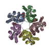
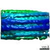
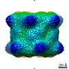
EMDB-28965:
Glutamine synthetase from Pseudomonas aeruginosa, filament double-unit in compressed conformation
Method: single particle / : Phan IQ, Staker B, Shek R, Moser TH, Evans JE, van Voorhis WC, Myler PJ, Seattle Structural Genomics Center for Infectious Disease (SSGCID)

PDB-8fbp:
Glutamine synthetase from Pseudomonas aeruginosa, filament double-unit in compressed conformation
Method: single particle / : Phan IQ, Staker B, Shek R, Moser TH, Evans JE, van Voorhis WC, Myler PJ, Seattle Structural Genomics Center for Infectious Disease (SSGCID)
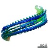
EMDB-10136:
Double-stranded helical ESCRT-III filament formed from Snf7/Vps24/Vps2 on a helical membrane bicelle
Method: helical / : Frost A, Johnson I, Talledge N
EMDB-10137:
Refined, asymmetrically masked double-stranded helical ESCRT-III filament formed from Snf7/Vps24/Vps2 on helical lipid bicelle
Method: helical / : Frost A, Johnson I, Talledge N
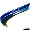
EMDB-10138:
Segment of helical membrane tube with longitudinal ESCRT-III filaments with different binding modes formed from Snf7/Vps24/Vps2
Method: subtomogram averaging / : Moser von Filseck J, Roux A
EMDB-10139:
Segment of helical membrane tube with longitudinal ESCRT-III filaments in the equatorial binding mode formed from Snf7/Vps24/Vps2
Method: subtomogram averaging / : Moser von Filseck J, Roux A
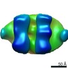
EMDB-4471:
Cryo-SOFI enabling low-dose super-resolution correlative light and electron cryo-microscopy
Method: electron tomography / : Moser F, Prazak V, Mordhorst V, Andrade AM, Baker LA, Hagen C, Grunewald K, Kaufmann R
EMDB-4472:
Cryo-SOFI enabling low-dose super-resolution correlative light and electron cryo-microscopy
Method: electron tomography / : Moser F, Prazak V, Mordhorst V, Andrade AM, Baker LA, Hagen C, Grunewald K, Kaufmann R
EMDB-4473:
Cryo-SOFI enabling low-dose super-resolution correlative light and electron cryo-microscopy
Method: electron tomography / : Moser F, Prazak V, Mordhorst V, Andrade AM, Baker LA, Hagen C, Grunewald K, Kaufmann R
EMDB-9190:
Cell-free-expressed Pyridoxal 5'-phosphate synthase-like subunit (PDX1.2) from Arabidopsis thaliana
Method: single particle / : Novikova IV, Evans JE, Hellmann H, Sharma N

PDB-6ez8:
Human Huntingtin-HAP40 complex structure
Method: single particle / : Guo Q, Bin H, Cheng J, Pfeifer G, Baumeister W, Fernandez-Busnadiego R, Kochanek S
 wwPDB to switch to version 3 of the EMDB data model
