 Movie
Movie Controller

[English] 日本語
 Yorodumi
- EMDB-4471: Cryo-SOFI enabling low-dose super-resolution correlative light an... -
+ Open data

- Basic information
| Entry | Database: EMDB / ID: EMD-4471 | |||||||||
|---|---|---|---|---|---|---|---|---|---|---|
| Title | Cryo-SOFI enabling low-dose super-resolution correlative light and electron cryo-microscopy | |||||||||
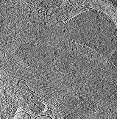
 Map data Map data | Tomogram of pseudopod of an XC cell. This cell was imaged with cryo-fluorescence microscopy super-resolution optical fluctiation imaging (cryoSOFI) prior to tilt-series acquisition. | |||||||||
 Sample Sample |
| |||||||||
| Biological species |  | |||||||||
| Method | electron tomography / cryo EM | |||||||||
 Authors Authors | Moser F / Prazak V / Mordhorst V / Andrade AM / Baker LA / Hagen C / Grunewald K / Kaufmann R | |||||||||
 Citation Citation | Journal: Proc Natl Acad Sci U S A / Year: 2019 Title: Cryo-SOFI enabling low-dose super-resolution correlative light and electron cryo-microscopy. Authors: Felipe Moser / Vojtěch Pražák / Valerie Mordhorst / Débora M Andrade / Lindsay A Baker / Christoph Hagen / Kay Grünewald / Rainer Kaufmann /   Abstract: Correlative light and electron cryo-microscopy (cryo-CLEM) combines information from the specific labeling of fluorescence cryo-microscopy (cryo-FM) with the high resolution in environmental context ...Correlative light and electron cryo-microscopy (cryo-CLEM) combines information from the specific labeling of fluorescence cryo-microscopy (cryo-FM) with the high resolution in environmental context of electron cryo-microscopy (cryo-EM). Exploiting super-resolution methods for cryo-FM is advantageous, as it enables the identification of rare events within the environmental background of cryo-EM at a sensitivity and resolution beyond that of conventional methods. However, due to the need for relatively high laser intensities, current super-resolution cryo-CLEM methods require cryo-protectants or support films which can severely reduce image quality in cryo-EM and are not compatible with many samples, such as mammalian cells. Here, we introduce cryogenic super-resolution optical fluctuation imaging (cryo-SOFI), a low-dose super-resolution imaging scheme based on the SOFI principle. As cryo-SOFI does not require special sample preparation, it is fully compatible with conventional cryo-EM specimens, and importantly, it does not affect the quality of cryo-EM imaging. By applying cryo-SOFI to a variety of biological application examples, we demonstrate resolutions up to ∼135 nm, an improvement of up to three times compared with conventional cryo-FM, while maintaining the specimen in a vitrified state for subsequent cryo-EM. Cryo-SOFI presents a general solution to the problem of specimen devitrification in super-resolution cryo-CLEM. It does not require a complex optical setup and can easily be implemented in any existing cryo-FM system. | |||||||||
| History |
|
- Structure visualization
| Movie |
 Movie viewer Movie viewer |
|---|---|
| Supplemental images |
- Downloads & links
-EMDB archive
| Map data | emd_4471.map.gz | 321.5 MB | EMDB map data format | |
|---|---|---|---|---|
| Header (meta data) | emd-4471-v30.xmlemd-4471.xml | 10.4 KB 10.4 KB | Display Display | EMDB header |
| Images |  emd_4471.png emd_4471.png | 303.3 KB | ||
| Archive directory |  http://ftp.pdbj.org/pub/emdb/structures/EMD-4471ftp://ftp.pdbj.org/pub/emdb/structures/EMD-4471 http://ftp.pdbj.org/pub/emdb/structures/EMD-4471ftp://ftp.pdbj.org/pub/emdb/structures/EMD-4471 | HTTPS FTP |
-Related structure data
-Links
| EMDB pages | EMDB (EBI/PDBe) / EMDataResource |
|---|
-Map
| File | Download / File: emd_4471.map.gz / Format: CCP4 / Size: 362.4 MB / Type: IMAGE STORED AS SIGNED INTEGER (2 BYTES) | ||||||||||||||||||||||||||||||||||||||||||||||||||||||||||||
|---|---|---|---|---|---|---|---|---|---|---|---|---|---|---|---|---|---|---|---|---|---|---|---|---|---|---|---|---|---|---|---|---|---|---|---|---|---|---|---|---|---|---|---|---|---|---|---|---|---|---|---|---|---|---|---|---|---|---|---|---|---|
| Annotation | Tomogram of pseudopod of an XC cell. This cell was imaged with cryo-fluorescence microscopy super-resolution optical fluctiation imaging (cryoSOFI) prior to tilt-series acquisition. | ||||||||||||||||||||||||||||||||||||||||||||||||||||||||||||
| Projections & slices | Image control
Images are generated by Spider. generated in cubic-lattice coordinate | ||||||||||||||||||||||||||||||||||||||||||||||||||||||||||||
| Voxel size | X=Y=Z: 16.88 Å | ||||||||||||||||||||||||||||||||||||||||||||||||||||||||||||
| Density |
| ||||||||||||||||||||||||||||||||||||||||||||||||||||||||||||
| Symmetry | Space group: 1 | ||||||||||||||||||||||||||||||||||||||||||||||||||||||||||||
| Details | EMDB XML:
CCP4 map header:
| ||||||||||||||||||||||||||||||||||||||||||||||||||||||||||||
 Z (Sec.)
 Y (Row.)
 X (Col.)


-Supplemental data
- Sample components
-Entire : XC (rous sarcoma) cell transiently expressing mClover-TOM20
| Entire | Name: XC (rous sarcoma) cell transiently expressing mClover-TOM20 |
|---|---|
| Components |
|
-Supramolecule #1: XC (rous sarcoma) cell transiently expressing mClover-TOM20
| Supramolecule | Name: XC (rous sarcoma) cell transiently expressing mClover-TOM20 type: cell / ID: 1 / Parent: 0 |
|---|---|
| Source (natural) | Organism: |
-Experimental details
-Structure determination
| Method | cryo EM |
|---|---|
 Processing Processing | electron tomography |
| Aggregation state | cell |
-Sample preparation
| Buffer | pH: 7.4 / Details: DMEM 10% fetal bofine serum |
|---|---|
| Grid | Material: GOLD / Mesh: 200 / Support film - #0 - Film type ID: 1 / Support film - #0 - Material: CARBON / Support film - #0 - topology: LACEY / Support film - #1 - Film type ID: 2 / Support film - #1 - Material: GRAPHENE OXIDE / Pretreatment - Type: GLOW DISCHARGE / Pretreatment - Atmosphere: AIR |
| Vitrification | Cryogen name: ETHANE-PROPANE / Instrument: HOMEMADE PLUNGER |
| Details | XC (rous sarcoma) cell transiently expressing mClover-TOM20 |
| Sectioning | Other: NO SECTIONING |
| Fiducial marker | Manufacturer: Aurion / Diameter: 10 nm |
- Electron microscopy
| Microscope | FEI POLARA 300 |
|---|---|
| Specialist optics | Energy filter - Slit width: 20 eV |
| Image recording | Film or detector model: GATAN K2 SUMMIT (4k x 4k) / Detector mode: COUNTING / Digitization - Dimensions - Width: 3836 pixel / Digitization - Dimensions - Height: 3710 pixel / Digitization - Frames/image: 1-20 / Average electron dose: 3.0 e/Å2 |
| Electron beam | Acceleration voltage: 300 kV / Electron source:  FIELD EMISSION GUN FIELD EMISSION GUN |
| Electron optics | C2 aperture diameter: 50.0 µm / Calibrated defocus max: 6.03 µm / Calibrated defocus min: 2.3 µm / Illumination mode: FLOOD BEAM / Imaging mode: BRIGHT FIELD / Cs: 2.0 mm / Nominal magnification: 50000 |
| Sample stage | Cooling holder cryogen: NITROGEN |
| Experimental equipment |  Model: Tecnai Polara / Image courtesy: FEI Company |
-Image processing
| Final reconstruction | Algorithm: BACK PROJECTION / Software - Name: eTomo / Number images used: 31 |
|---|---|
| CTF correction | Software - Name: CTFPHASEFLIP |
