 ムービー
ムービー コントローラー
 構造ビューア
 EMN検索について


-検索条件
-検索結果
検索 (著者・登録者: may & a)の結果1,066件中、1から50件目までを表示しています

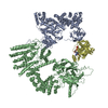
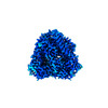
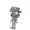
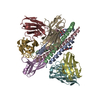
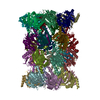
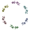
EMDB-38466: 
Cryo-EM structure of the RhoG/DOCK5/ELMO1/Rac1 complex: RhoG/DOCK5/ELMO1 focused map

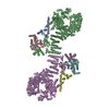
EMDB-60136:
Cryo-EM structure of the RhoG/DOCK5/ELMO1/Rac1 complex

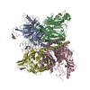
EMDB-60146:
Structure of DOCK5/ELMO1/Rac1 core (RhoG/DOCK5/ELMO1/Rac1 dataset, class 1)

EMDB-60147:
Structure of DOCK5/ELMO1/Rac1 core (RhoG/DOCK5/ELMO1/Rac1 dataset, class 2)

EMDB-60148:
Structure of DOCK5/ELMO1/Rac1 core (RhoG/DOCK5/ELMO1/Rac1 dataset, class 3)

EMDB-60149:
Structure of DOCK5/ELMO1/Rac1 core (RhoG/DOCK5/ELMO1/Rac1 dataset, class 4)

EMDB-60150:
Structure of DOCK5/ELMO1/Rac1 core (RhoG/DOCK5/ELMO1/Rac1 dataset, class 5)
PDB-8xm7:
Cryo-EM structure of the RhoG/DOCK5/ELMO1/Rac1 complex: RhoG/DOCK5/ELMO1 focused map

PDB-8zj2:
Cryo-EM structure of the RhoG/DOCK5/ELMO1/Rac1 complex

PDB-8zji:
Structure of DOCK5/ELMO1/Rac1 core (RhoG/DOCK5/ELMO1/Rac1 dataset, class 1)
PDB-8zjj:
Structure of DOCK5/ELMO1/Rac1 core (RhoG/DOCK5/ELMO1/Rac1 dataset, class 2)
PDB-8zjk:
Structure of DOCK5/ELMO1/Rac1 core (RhoG/DOCK5/ELMO1/Rac1 dataset, class 3)
PDB-8zjl:
Structure of DOCK5/ELMO1/Rac1 core (RhoG/DOCK5/ELMO1/Rac1 dataset, class 4)

PDB-8zjm:
Structure of DOCK5/ELMO1/Rac1 core (RhoG/DOCK5/ELMO1/Rac1 dataset, class 5)

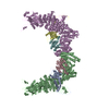
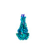
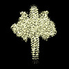
EMDB-42770:
Caulobacter crescentus FljM flagellar filament (symmetrized)
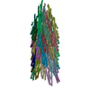
PDB-8uxn:
Caulobacter crescentus FljM flagellar filament (symmetrized)

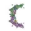
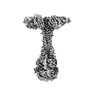
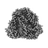
EMDB-42766:
Caulobacter crescentus FljK flagellar filament (asymmetrical)
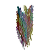
PDB-8uxj:
Caulobacter crescentus FljK flagellar filament (asymmetrical)

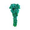
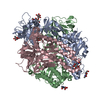
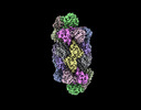
EMDB-36271:
Cryo-EM structure of the DOCK5/ELMO1 complex, focused on one protomer

PDB-8jhk:
Cryo-EM structure of the DOCK5/ELMO1 complex, focused on one protomer
EMDB-41639:
Langya henipavirus fusion protein in postfusion state
EMDB-41640:
Langya henipavirus fusion protein in prefusion state
EMDB-41641:
Langya henipavirus postfusion F protein in complex with the 4G5 Fab, local refinement of the viral membrane distal region

EMDB-41642:
Langya henipavirus postfusion F protein in complex with 4G5 Fab, local refinement of the viral membrane proximal region
EMDB-41644:
Langya henipavirus postfusion fusion protein in complex with 4G5 Fab (global refinement)

PDB-8tve:
Langya henipavirus fusion protein in postfusion state
PDB-8tvf:
Langya henipavirus fusion protein in prefusion state
PDB-8tvg:
Langya henipavirus postfusion F protein in complex with the 4G5 Fab, local refinement of the viral membrane distal region
PDB-8tvh:
Langya henipavirus postfusion F protein in complex with 4G5 Fab, local refinement of the viral membrane proximal region
EMDB-41636:
Ghanaian virus fusion glycoprotein (GhV F)
EMDB-41643:
Langya Virus G glycoprotein (LayV G) with stabilizing mutations

EMDB-43593:
Langya Virus attachment (G) glycoprotein with K85L/L86K mutation

PDB-8tvb:
Ghanaian virus fusion glycoprotein (GhV F)

PDB-8tvi:
Langya Virus G glycoprotein (LayV G) with stabilizing mutations
PDB-8vwp:
Langya Virus attachment (G) glycoprotein with K85L/L86K mutation
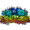
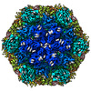
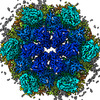
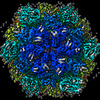
EMDB-16489:
In situ structure of the Nitrosopumilus maritimus S-layer - Six-fold symmetry (C6)
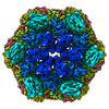
EMDB-16492:
In situ structure of the Nitrosopumilus maritimus S-layer - Composite map between C2 and C6

PDB-8c8o:
In situ structure of the Nitrosopumilus maritimus S-layer - Six-fold symmetry (C6)
PDB-8c8r:
In situ structure of the Nitrosopumilus maritimus S-layer - Composite map between C2 and C6
EMDB-41963:
Preholo-Proteasome from Beta 3 D205 deletion

EMDB-41993:
Proteasome 20S Core Particle from Beta 3 D205 deletion

PDB-8u6y:
Preholo-Proteasome from Beta 3 D205 deletion
PDB-8u7u:
Proteasome 20S Core Particle from Beta 3 D205 deletion

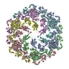
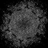
EMDB-16482:
In vitro structure of the Nitrosopumilus maritimus S-layer - Six-fold symmetry (C6)
EMDB-16483:
In vitro structure of the Nitrosopumilus maritimus S-layer - Two-fold symmetry (C2)
EMDB-16484:
In vitro structure of the Nitrosopumilus maritimus S-layer - Composite map between two and six-fold symmetrised
EMDB-16486:
In vitro Nitrosopumilus maritimus S-layer with NH4Cl
EMDB-16487:
In situ structure of the Nitrosopumilus maritimus S-layer - Two-fold symmetry (C2)
PDB-8c8k:
In vitro structure of the Nitrosopumilus maritimus S-layer - Six-fold symmetry (C6)

PDB-8c8l:
In vitro structure of the Nitrosopumilus maritimus S-layer - Two-fold symmetry (C2)
ページ:
 wwPDBはEMDBデータモデルのバージョン3へ移行します
