 Movie
Movie Controller


+ Open data

- Basic information
| Entry | Database: PDB / ID: 8u6y | ||||||
|---|---|---|---|---|---|---|---|
| Title | Preholo-Proteasome from Beta 3 D205 deletion | ||||||
 Components Components |
| ||||||
 Keywords Keywords | HYDROLASE / Proteasome / core particle | ||||||
| Function / homology |  Function and homology information Function and homology informationER-Phagosome pathway / Antigen processing: Ub, ATP-independent proteasomal degradation / proteasome core complex assembly / Proteasome assembly / Cross-presentation of soluble exogenous antigens (endosomes) / TNFR2 non-canonical NF-kB pathway / nuclear outer membrane-endoplasmic reticulum membrane network / Regulation of PTEN stability and activity / CDK-mediated phosphorylation and removal of Cdc6 / FBXL7 down-regulates AURKA during mitotic entry and in early mitosis ...ER-Phagosome pathway / Antigen processing: Ub, ATP-independent proteasomal degradation / proteasome core complex assembly / Proteasome assembly / Cross-presentation of soluble exogenous antigens (endosomes) / TNFR2 non-canonical NF-kB pathway / nuclear outer membrane-endoplasmic reticulum membrane network / Regulation of PTEN stability and activity / CDK-mediated phosphorylation and removal of Cdc6 / FBXL7 down-regulates AURKA during mitotic entry and in early mitosis / KEAP1-NFE2L2 pathway / Neddylation / Ubiquitin-Mediated Degradation of Phosphorylated Cdc25A / Orc1 removal from chromatin / MAPK6/MAPK4 signaling / Antigen processing: Ubiquitination & Proteasome degradation / proteasome storage granule / proteasomal ubiquitin-independent protein catabolic process / Ub-specific processing proteases / proteasome endopeptidase complex / proteasome core complex, beta-subunit complex / endopeptidase activator activity / threonine-type endopeptidase activity / proteasome assembly / proteasome core complex, alpha-subunit complex / Neutrophil degranulation / proteasome complex / peroxisome / endopeptidase activity / ubiquitin-dependent protein catabolic process / proteasome-mediated ubiquitin-dependent protein catabolic process / mRNA binding / DNA damage response / endoplasmic reticulum membrane / mitochondrion / nucleus / cytoplasm / cytosol Similarity search - Function | ||||||
| Biological species |  | ||||||
| Method | ELECTRON MICROSCOPY / single particle reconstruction / cryo EM / Resolution: 2.8 Å | ||||||
 Authors Authors | Walsh Jr., R.M. / Rawson, S. / Velez, B. / Blickling, M. / Razi, A. / Hanna, J. | ||||||
| Funding support |  United States, 1items United States, 1items
| ||||||
 Citation Citation | Journal: Nat Struct Mol Biol / Year: 2024 Title: Mechanism of autocatalytic activation during proteasome assembly. Authors: Benjamin Velez / Richard M Walsh / Shaun Rawson / Aida Razi / Lea Adams / Erignacio Fermin Perez / Fenglong Jiao / Marie Blickling / Tamayanthi Rajakumar / Darlene Fung / Lan Huang / John Hanna / Abstract: Many large molecular machines are too elaborate to assemble spontaneously and are built through ordered pathways orchestrated by dedicated chaperones. During assembly of the core particle (CP) of the ...Many large molecular machines are too elaborate to assemble spontaneously and are built through ordered pathways orchestrated by dedicated chaperones. During assembly of the core particle (CP) of the proteasome, where protein degradation occurs, its six active sites are simultaneously activated via cleavage of N-terminal propeptides. Such activation is autocatalytic and coupled to fusion of two half-CP intermediates, which protects cells by preventing activation until enclosure of the active sites within the CP interior. Here we uncover key mechanistic aspects of autocatalytic activation, which proceeds through alignment of the β5 and β2 catalytic triad residues, respectively, with these triads being misaligned before fusion. This mechanism contrasts with most other zymogens, in which catalytic centers are preformed. Our data also clarify the mechanism by which individual subunits can be added in a precise, temporally ordered manner. This work informs two decades-old mysteries in the proteasome field, with broader implications for protease biology and multisubunit complex assembly. | ||||||
| History |
|
- Structure visualization
| Structure viewer | Molecule: MolmilJmol/JSmol |
|---|
- Downloads & links
-Download
| PDBx/mmCIF format | 8u6y.cif.gz | 2.6 MB | Display | PDBx/mmCIF format |
|---|---|---|---|---|
| PDB format | pdb8u6y.ent.gz | Display | PDB format | |
| PDBx/mmJSON format | 8u6y.json.gz | Tree view | PDBx/mmJSON format | |
| Others |  Other downloads Other downloads |
-Validation report
| Arichive directory | https://data.pdbj.org/pub/pdb/validation_reports/u6/8u6yftp://data.pdbj.org/pub/pdb/validation_reports/u6/8u6y | HTTPS FTP |
|---|
-Related structure data
| Related structure data |  41963MC 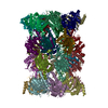 8u7uC M: map data used to model this data C: citing same article ( |
|---|---|
| Similar structure data |

-Links
 PDBj
PDBj


- Assembly
| Deposited unit | 
|
|---|---|
| 1 |
|
-Components
-Proteasome subunit alpha type- ... , 7 types, 14 molecules ARBSCTDUEVFWGX
| #1: Protein | Mass: 28033.830 Da / Num. of mol.: 2 / Source method: isolated from a natural source / Source: (natural) References: UniProt: P21243, proteasome endopeptidase complex #2: Protein | Mass: 27191.828 Da / Num. of mol.: 2 / Source method: isolated from a natural source / Source: (natural) References: UniProt: P23639, proteasome endopeptidase complex #3: Protein | Mass: 27181.609 Da / Num. of mol.: 2 / Source method: isolated from a natural source / Source: (natural) #4: Protein | Mass: 28478.111 Da / Num. of mol.: 2 / Source method: isolated from a natural source / Source: (natural) References: UniProt: P40303, proteasome endopeptidase complex #5: Protein | Mass: 28649.086 Da / Num. of mol.: 2 / Source method: isolated from a natural source / Source: (natural) References: UniProt: P32379, proteasome endopeptidase complex #6: Protein | Mass: 25634.000 Da / Num. of mol.: 2 / Source method: isolated from a natural source / Source: (natural) References: UniProt: P40302, proteasome endopeptidase complex #7: Protein | Mass: 31575.068 Da / Num. of mol.: 2 / Source method: isolated from a natural source / Source: (natural) References: UniProt: P21242, proteasome endopeptidase complex |
|---|
-Protein , 3 types, 6 molecules HYOfPg
| #8: Protein | Mass: 16777.766 Da / Num. of mol.: 2 / Source method: isolated from a natural source / Source: (natural) #15: Protein | Mass: 30718.074 Da / Num. of mol.: 2 / Source method: isolated from a natural source / Source: (natural) #16: Protein | Mass: 30762.895 Da / Num. of mol.: 2 / Source method: isolated from a natural source / Source: (natural) |
|---|
-Proteasome subunit beta type- ... , 7 types, 14 molecules IZJaKbLcMdNeQh
| #9: Protein | Mass: 28299.889 Da / Num. of mol.: 2 / Source method: isolated from a natural source / Source: (natural) References: UniProt: P25043, proteasome endopeptidase complex #10: Protein | Mass: 22512.754 Da / Num. of mol.: 2 / Source method: isolated from a natural source / Source: (natural) #11: Protein | Mass: 22545.676 Da / Num. of mol.: 2 / Source method: isolated from a natural source / Source: (natural) References: UniProt: P22141, proteasome endopeptidase complex #12: Protein | Mass: 31670.539 Da / Num. of mol.: 2 / Source method: isolated from a natural source / Source: (natural) References: UniProt: P30656, proteasome endopeptidase complex #13: Protein | Mass: 26905.076 Da / Num. of mol.: 2 / Source method: isolated from a natural source / Source: (natural) References: UniProt: P23724, proteasome endopeptidase complex #14: Protein | Mass: 23573.604 Da / Num. of mol.: 2 / Source method: isolated from a natural source / Source: (natural) References: UniProt: P38624, proteasome endopeptidase complex #17: Protein | Mass: 29471.289 Da / Num. of mol.: 2 / Source method: isolated from a natural source / Source: (natural) References: UniProt: P30657, proteasome endopeptidase complex |
|---|
-Details
| Has protein modification | Y |
|---|
-Experimental details
-Experiment
| Experiment | Method: ELECTRON MICROSCOPY |
|---|---|
| EM experiment | Aggregation state: PARTICLE / 3D reconstruction method: single particle reconstruction |
- Sample preparation
| Component | Name: Preholo-proteasome from Beta 3 D205 deletion mutant / Type: COMPLEX Details: D205 is terminal most residue in the Beta 3 subunit. The deletion of this residue causes an accumulation of assembly intermediates including the Preholo-Proteasome Entity ID: all / Source: NATURAL | ||||||||||||||||||||
|---|---|---|---|---|---|---|---|---|---|---|---|---|---|---|---|---|---|---|---|---|---|
| Molecular weight | Value: 0.75 MDa / Experimental value: NO | ||||||||||||||||||||
| Source (natural) | Organism: | ||||||||||||||||||||
| Buffer solution | pH: 7.5 Details: Fluorinated Fos-Choline was added immediately prior to deposition on a grid for plunge freezing. | ||||||||||||||||||||
| Buffer component |
| ||||||||||||||||||||
| Specimen | Conc.: 4.48 mg/ml / Embedding applied: NO / Shadowing applied: NO / Staining applied: NO / Vitrification applied: YES | ||||||||||||||||||||
| Specimen support | Grid material: COPPER / Grid mesh size: 400 divisions/in. / Grid type: Quantifoil R1.2/1.3 | ||||||||||||||||||||
| Vitrification | Instrument: FEI VITROBOT MARK IV / Cryogen name: ETHANE / Humidity: 100 % / Chamber temperature: 295.15 K |
- Electron microscopy imaging
| Experimental equipment |  Model: Titan Krios / Image courtesy: FEI Company |
|---|---|
| Microscopy | Model: FEI TITAN KRIOS |
| Electron gun | Electron source:  FIELD EMISSION GUN / Accelerating voltage: 300 kV / Illumination mode: FLOOD BEAM FIELD EMISSION GUN / Accelerating voltage: 300 kV / Illumination mode: FLOOD BEAM |
| Electron lens | Mode: BRIGHT FIELD / Nominal magnification: 81000 X / Calibrated magnification: 47169 X / Nominal defocus max: 2200 nm / Nominal defocus min: 800 nm / Cs: 2.7 mm / C2 aperture diameter: 50 µm / Alignment procedure: COMA FREE |
| Specimen holder | Cryogen: NITROGEN / Specimen holder model: FEI TITAN KRIOS AUTOGRID HOLDER |
| Image recording | Average exposure time: 3.995 sec. / Electron dose: 57 e/Å2 / Film or detector model: GATAN K3 BIOQUANTUM (6k x 4k) / Num. of grids imaged: 1 / Num. of real images: 7461 |
| EM imaging optics | Energyfilter name: GIF Bioquantum / Energyfilter slit width: 25 eV |
- Processing
| EM software |
| |||||||||||||||||||||||||||||||||||||||||||||||||||||||
|---|---|---|---|---|---|---|---|---|---|---|---|---|---|---|---|---|---|---|---|---|---|---|---|---|---|---|---|---|---|---|---|---|---|---|---|---|---|---|---|---|---|---|---|---|---|---|---|---|---|---|---|---|---|---|---|---|
| CTF correction | Type: PHASE FLIPPING AND AMPLITUDE CORRECTION | |||||||||||||||||||||||||||||||||||||||||||||||||||||||
| Particle selection | Num. of particles selected: 1146597 | |||||||||||||||||||||||||||||||||||||||||||||||||||||||
| Symmetry | Point symmetry: C2 (2 fold cyclic) | |||||||||||||||||||||||||||||||||||||||||||||||||||||||
| 3D reconstruction | Resolution: 2.8 Å / Resolution method: FSC 0.143 CUT-OFF / Num. of particles: 19020 / Symmetry type: POINT | |||||||||||||||||||||||||||||||||||||||||||||||||||||||
| Atomic model building | Protocol: AB INITIO MODEL / Space: REAL | |||||||||||||||||||||||||||||||||||||||||||||||||||||||
| Atomic model building | PDB-ID: 8T08 Accession code: 8T08 / Source name: PDB / Type: experimental model |

