 ムービー
ムービー コントローラー
 構造ビューア
 EMN検索について


-検索条件
-検索結果
検索 (著者・登録者: elisa & o)の結果533件中、1から50件目までを表示しています
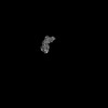
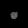
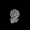
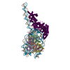
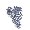
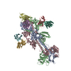
EMDB-43813: 
VIR-7229 Fab fragment bound the SARS-CoV-2 BA.2.86 spike trimer (local refinement of the BA 2.86 RBD/VIR-7229 VHVL)
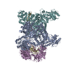
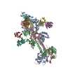
EMDB-43842:
VIR-7229 Fab fragment bound the BA.2.86 spike trimer (global refinement)

PDB-9asd:
VIR-7229 Fab fragment bound the SARS-CoV-2 BA.2.86 spike trimer (local refinement of the BA 2.86 RBD/VIR-7229 VHVL)
PDB-9au2:
VIR-7229 Fab fragment bound the BA.2.86 spike trimer (global refinement)
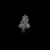
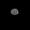
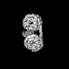
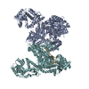
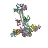
EMDB-51238:
Structure of a hexasome-nucleosome complex with a dyad-to-dyad distance of 103 bp.
EMDB-51239:
Nucleosome portion of SHN103, unsharpened focused refinement.
EMDB-51240:
Hexasome portion of SHN103, unsharpened focused refinement.
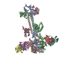
EMDB-51241:
Structure of Chd1 bound to a hexasome-nucleosome complex with a dyad-to-dyad distance of 103 bp.
EMDB-51242:
Nucleosome portion of Chd1-bound SHN103, unsharpened focused refinement.
EMDB-51243:
Hexasome portion of Chd1-bound SHN103, unsharpened focused refinement.
EMDB-51244:
Structure of Chd1 bound to a dinucleosome with a dyad-to-dyad distance of 103 bp.
EMDB-51245:
Original nucleosome portion of DN103, unsharpened focused refinement
EMDB-51246:
Restored Chd1-bound nucleosome portion of DN103, unsharpened focused refinement
EMDB-51247:
Structure of a mononucleosome bound by one copy of Chd1 with the DBD on the exit-side DNA.

EMDB-51315:
Unsharpened consensus map of hexasome-nucleosome complex SHN103

EMDB-51316:
Unsharpened consensus map of hexasome-nucleosome complex SHN103 bound by Chd1
EMDB-51317:
Unsharpened consensus map of dinucleosome DN103 bound by Chd1
PDB-9gd0:
Structure of a hexasome-nucleosome complex with a dyad-to-dyad distance of 103 bp.

PDB-9gd1:
Structure of Chd1 bound to a hexasome-nucleosome complex with a dyad-to-dyad distance of 103 bp.
PDB-9gd2:
Structure of Chd1 bound to a dinucleosome with a dyad-to-dyad distance of 103 bp.
PDB-9gd3:
Structure of a mononucleosome bound by one copy of Chd1 with the DBD on the exit-side DNA.

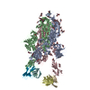
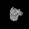
EMDB-41785:
DdmD dimer in complex with ssDNA
EMDB-41790:
DdmD monomer in complex with ssDNA
EMDB-41865:
DdmDE handover complex
PDB-8u0u:
DdmD dimer in complex with ssDNA
PDB-8u0w:
DdmD monomer in complex with ssDNA
PDB-8u3k:
DdmDE handover complex
EMDB-41781:
DdmE in complex with guide and target DNA
EMDB-44825:
DdmD dimer apoprotein (CASP target)
PDB-8u0j:
DdmE in complex with guide and target DNA
PDB-9bqv:
DdmD dimer apoprotein
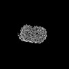
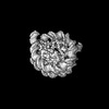
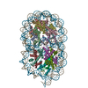
EMDB-19163:
Trimeric HSV-1F gB ectodomain in postfusion conformation with three bound HDIT101 Fab molecules.

EMDB-19164:
Trimeric HSV-1F gB ectodomain in postfusion conformation with three bound HDIT102 Fab molecules.

EMDB-19165:
Trimeric HSV-2F gB ectodomain in postfusion conformation with three bound HDIT101 Fab molecules.
EMDB-19166:
Trimeric HSV-2G gB ectodomain in postfusion conformation with three bound HDIT102 Fab molecules.
PDB-8rgz:
Trimeric HSV-1F gB ectodomain in postfusion conformation with three bound HDIT101 Fab molecules.
PDB-8rh0:
Trimeric HSV-1F gB ectodomain in postfusion conformation with three bound HDIT102 Fab molecules.
PDB-8rh1:
Trimeric HSV-2F gB ectodomain in postfusion conformation with three bound HDIT101 Fab molecules.
PDB-8rh2:
Trimeric HSV-2G gB ectodomain in postfusion conformation with three bound HDIT102 Fab molecules.

EMDB-17377:
Structure of human SIT1 (focussed map / refinement)

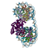
EMDB-17378:
Structure of human SIT1:ACE2 complex (open PD conformation)

EMDB-17379:
Structure of human SIT1:ACE2 complex (closed PD conformation)

EMDB-17380:
Structure of human SIT1 bound to L-pipecolate (focussed map / refinement)

EMDB-17381:
Structure of human SIT1:ACE2 complex (open PD conformation) bound to L-pipecolate

EMDB-17382:
Structure of human SIT1:ACE2 complex (closed PD conformation) bound to L-pipecolate

PDB-8p2w:
Structure of human SIT1 (focussed map / refinement)

PDB-8p2x:
Structure of human SIT1:ACE2 complex (open PD conformation)

PDB-8p2y:
Structure of human SIT1:ACE2 complex (closed PD conformation)

PDB-8p2z:
Structure of human SIT1 bound to L-pipecolate (focussed map / refinement)
PDB-8p30:
Structure of human SIT1:ACE2 complex (open PD conformation) bound to L-pipecolate
ページ:
 wwPDBはEMDBデータモデルのバージョン3へ移行します
