[English] 日本語
 Yorodumi
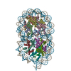
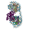
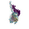
- EMDB-51242: Nucleosome portion of Chd1-bound SHN103, unsharpened focused refi... -
+ Open data
- Basic information
| Entry |  | |||||||||
|---|---|---|---|---|---|---|---|---|---|---|
| Title | Nucleosome portion of Chd1-bound SHN103, unsharpened focused refinement. | |||||||||
 Map data Map data | Nucleosome portion of Chd1-bound SHN103 | |||||||||
 Sample Sample |
| |||||||||
 Keywords Keywords | chromatin / remodeling / transcription / nucleosome / DNA BINDING PROTEIN | |||||||||
| Biological species |  | |||||||||
| Method | single particle reconstruction / cryo EM / Resolution: 4.0 Å | |||||||||
 Authors Authors | Engeholm M / Roske JJ / Oberbeckmann E / Dienemann C / Lidschreiber M / Cramer P / Farnung L | |||||||||
| Funding support |  Germany, European Union, 2 items Germany, European Union, 2 items
| |||||||||
 Citation Citation |  Journal: Mol Cell / Year: 2024 Journal: Mol Cell / Year: 2024Title: Resolution of transcription-induced hexasome-nucleosome complexes by Chd1 and FACT. Authors: Maik Engeholm / Johann J Roske / Elisa Oberbeckmann / Christian Dienemann / Michael Lidschreiber / Patrick Cramer / Lucas Farnung /    Abstract: To maintain the nucleosome organization of transcribed genes, ATP-dependent chromatin remodelers collaborate with histone chaperones. Here, we show that at the 5' ends of yeast genes, RNA polymerase ...To maintain the nucleosome organization of transcribed genes, ATP-dependent chromatin remodelers collaborate with histone chaperones. Here, we show that at the 5' ends of yeast genes, RNA polymerase II (RNAPII) generates hexasomes that occur directly adjacent to nucleosomes. The resulting hexasome-nucleosome complexes are then resolved by Chd1. We present two cryoelectron microscopy (cryo-EM) structures of Chd1 bound to a hexasome-nucleosome complex before and after restoration of the missing inner H2A/H2B dimer by FACT. Chd1 uniquely interacts with the complex, positioning its ATPase domain to shift the hexasome away from the nucleosome. In the absence of the inner H2A/H2B dimer, its DNA-binding domain (DBD) packs against the ATPase domain, suggesting an inhibited state. Restoration of the dimer by FACT triggers a rearrangement that displaces the DBD and stimulates Chd1 remodeling. Our results demonstrate how chromatin remodelers interact with a complex nucleosome assembly and suggest how Chd1 and FACT jointly support transcription by RNAPII. | |||||||||
| History |
|
- Structure visualization
| Supplemental images |
|---|
- Downloads & links
-EMDB archive
| Map data |  emd_51242.map.gz emd_51242.map.gz | 65.2 MB |  EMDB map data format EMDB map data format | |
|---|---|---|---|---|
| Header (meta data) |  emd-51242-v30.xml emd-51242-v30.xml emd-51242.xml emd-51242.xml | 13.3 KB 13.3 KB | Display Display |  EMDB header EMDB header |
| FSC (resolution estimation) |  emd_51242_fsc.xml emd_51242_fsc.xml | 10 KB | Display |  FSC data file FSC data file |
| Images |  emd_51242.png emd_51242.png | 39.3 KB | ||
| Masks |  emd_51242_msk_1.map emd_51242_msk_1.map | 83.7 MB |  Mask map Mask map | |
| Filedesc metadata |  emd-51242.cif.gz emd-51242.cif.gz | 3.9 KB | ||
| Others |  emd_51242_half_map_1.map.gz emd_51242_half_map_1.map.gz emd_51242_half_map_2.map.gz emd_51242_half_map_2.map.gz | 65.2 MB 65.3 MB | ||
| Archive directory |  http://ftp.pdbj.org/pub/emdb/structures/EMD-51242 http://ftp.pdbj.org/pub/emdb/structures/EMD-51242 ftp://ftp.pdbj.org/pub/emdb/structures/EMD-51242 ftp://ftp.pdbj.org/pub/emdb/structures/EMD-51242 | HTTPS FTP |
-Validation report
| Summary document |  emd_51242_validation.pdf.gz emd_51242_validation.pdf.gz | 900 KB | Display |  EMDB validaton report EMDB validaton report |
|---|---|---|---|---|
| Full document |  emd_51242_full_validation.pdf.gz emd_51242_full_validation.pdf.gz | 899.5 KB | Display | |
| Data in XML |  emd_51242_validation.xml.gz emd_51242_validation.xml.gz | 16.9 KB | Display | |
| Data in CIF |  emd_51242_validation.cif.gz emd_51242_validation.cif.gz | 22.1 KB | Display | |
| Arichive directory |  https://ftp.pdbj.org/pub/emdb/validation_reports/EMD-51242 https://ftp.pdbj.org/pub/emdb/validation_reports/EMD-51242 ftp://ftp.pdbj.org/pub/emdb/validation_reports/EMD-51242 ftp://ftp.pdbj.org/pub/emdb/validation_reports/EMD-51242 | HTTPS FTP |
-Related structure data
- Links
Links
| EMDB pages |  EMDB (EBI/PDBe) / EMDB (EBI/PDBe) /  EMDataResource EMDataResource |
|---|
- Map
Map
| File |  Download / File: emd_51242.map.gz / Format: CCP4 / Size: 83.7 MB / Type: IMAGE STORED AS FLOATING POINT NUMBER (4 BYTES) Download / File: emd_51242.map.gz / Format: CCP4 / Size: 83.7 MB / Type: IMAGE STORED AS FLOATING POINT NUMBER (4 BYTES) | ||||||||||||||||||||||||||||||||||||
|---|---|---|---|---|---|---|---|---|---|---|---|---|---|---|---|---|---|---|---|---|---|---|---|---|---|---|---|---|---|---|---|---|---|---|---|---|---|
| Annotation | Nucleosome portion of Chd1-bound SHN103 | ||||||||||||||||||||||||||||||||||||
| Projections & slices | Image control
Images are generated by Spider. | ||||||||||||||||||||||||||||||||||||
| Voxel size | X=Y=Z: 0.834 Å | ||||||||||||||||||||||||||||||||||||
| Density |
| ||||||||||||||||||||||||||||||||||||
| Symmetry | Space group: 1 | ||||||||||||||||||||||||||||||||||||
| Details | EMDB XML:
|
-Supplemental data
-Mask #1
| File |  emd_51242_msk_1.map emd_51242_msk_1.map | ||||||||||||
|---|---|---|---|---|---|---|---|---|---|---|---|---|---|
| Projections & Slices |
| ||||||||||||
| Density Histograms |
-Half map: #1
| File | emd_51242_half_map_1.map | ||||||||||||
|---|---|---|---|---|---|---|---|---|---|---|---|---|---|
| Projections & Slices |
| ||||||||||||
| Density Histograms |
-Half map: #2
| File | emd_51242_half_map_2.map | ||||||||||||
|---|---|---|---|---|---|---|---|---|---|---|---|---|---|
| Projections & Slices |
| ||||||||||||
| Density Histograms |
- Sample components
-Entire : Chd1 bound to a hexasome-nucleosome complex with a dyad-to-dyad d...
| Entire | Name: Chd1 bound to a hexasome-nucleosome complex with a dyad-to-dyad distance of 103 bp |
|---|---|
| Components |
|
-Supramolecule #1: Chd1 bound to a hexasome-nucleosome complex with a dyad-to-dyad d...
| Supramolecule | Name: Chd1 bound to a hexasome-nucleosome complex with a dyad-to-dyad distance of 103 bp type: complex / ID: 1 / Parent: 0 / Macromolecule list: #1-#7 |
|---|---|
| Source (natural) | Organism:  |
-Experimental details
-Structure determination
| Method | cryo EM |
|---|---|
 Processing Processing | single particle reconstruction |
| Aggregation state | particle |
- Sample preparation
| Buffer | pH: 7.4 |
|---|---|
| Vitrification | Cryogen name: ETHANE |
- Electron microscopy
| Microscope | FEI TITAN KRIOS |
|---|---|
| Image recording | Film or detector model: GATAN K3 (6k x 4k) / Average electron dose: 39.8 e/Å2 |
| Electron beam | Acceleration voltage: 300 kV / Electron source:  FIELD EMISSION GUN FIELD EMISSION GUN |
| Electron optics | Illumination mode: FLOOD BEAM / Imaging mode: BRIGHT FIELD / Nominal defocus max: 2.0 µm / Nominal defocus min: 0.5 µm |
| Experimental equipment |  Model: Titan Krios / Image courtesy: FEI Company |
 Movie
Movie Controller


 Z (Sec.)
 Y (Row.)
 X (Col.)


